Foltyn Martha, Nieto Taborda Karen Natalia, Neuberger Ulf, Brugnara Gianluca, Reinhardt Annekathrin, Stichel Damian, Heiland Sabine, Herold-Mende Christel, Unterberg Andreas, Debus Jürgen, von Deimling Andreas, Wick Wolfgang, Bendszus Martin, Kickingereder Philipp
Department of Neuroradiology, University of Heidelberg Medical Center, Heidelberg, Germany.
Department of Neuropathology, University of Heidelberg Medical Center, Heidelberg, Germany.
Neurooncol Adv. 2020 Jan 10;2(1):vdaa004. doi: 10.1093/noajnl/vdaa004. eCollection 2020 Jan-Dec.
This study aimed to assess the validity and pathophysiology of the T2/FLAIR-mismatch sign for noninvasive identification of isocitrate dehydrogenase (IDH)-mutant 1p/19q non-codeleted glioma.
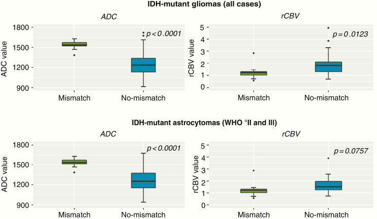
Magnetic resonance imaging scans from 408 consecutive patients with newly diagnosed glioma (113 lower-grade gliomas and 295 glioblastomas) were evaluated for the presence of T2/FLAIR-mismatch sign by 2 independent reviewers. Sensitivity, specificity, accuracy, positive predictive value (PPV), and negative predictive value (NPV) were calculated to assess the performance of the T2/FLAIR-mismatch sign for identifying IDH-mutant 1p/19q non-codeleted tumors. An exploratory analysis of differences in contrast-enhancing tumor volumes, apparent diffusion coefficient (ADC) values, and relative cerebral blood volume (rCBV) values in IDH-mutant gliomas with versus without the presence of a T2/FLAIR-mismatch sign (as well as analysis of spatial differences within tumors with the presence of a T2/FLAIR-mismatch sign) was performed.
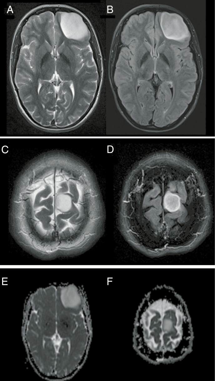
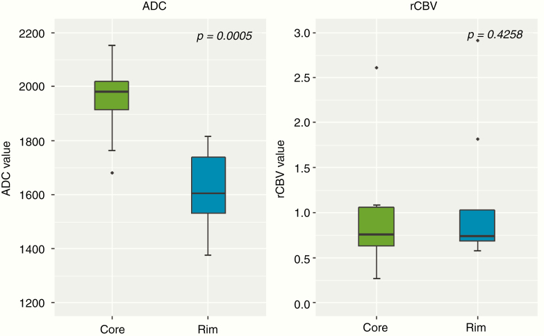
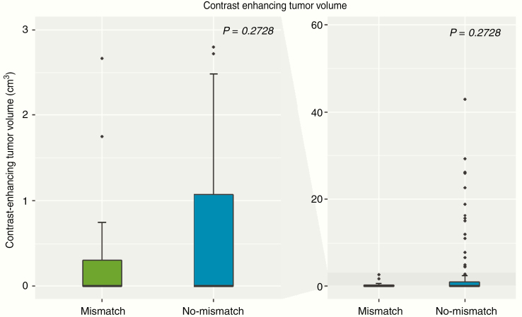
The T2/FLAIR-mismatch sign was present in 12 cases with lower-grade glioma (10.6%), all of them being IDH-mutant 1p/19q non-codeleted tumors (sensitivity = 10.9%, specificity = 100%, PPV = 100%, NPV = 3.0%, accuracy = 13.3%). There was a substantial interrater agreement to identify the T2/FLAIR-mismatch sign (Cohen's kappa = 0.75 [95% CI, 0.57-0.93]). The T2/FLAIR-mismatch sign was not identified in any other molecular subgroup, including IDH-mutant glioblastoma cases ( = 5). IDH-mutant gliomas with a T2/FLAIR-mismatch sign showed significantly higher ADC ( < .0001) and lower rCBV values ( = .0123) as compared to IDH-mutant gliomas without a T2/FLAIR-mismatch sign. Moreover, in IDH-mutant gliomas with T2/FLAIR-mismatch sign the ADC values were significantly lower in the FLAIR-hyperintense rim as compared to the FLAIR-hypointense core of the tumor ( = .0005).
This study confirms the high specificity of the T2/FLAIR-mismatch sign for noninvasive identification of IDH-mutant 1p/19q non-codeleted gliomas; however, sensitivity is low and applicability is limited to lower-grade gliomas. Whether the higher ADC and lower rCBV values in IDH-mutant gliomas with a T2/FLAIR-mismatch sign (as compared to those without) translate into a measurable prognostic effect requires investigation in future studies. Moreover, spatial differences in ADC values between the core and rim of tumors with a T2/FLAIR-mismatch sign potentially reflect specific distinctions in tumor cellularity and microenvironment.
本研究旨在评估T2/液体衰减反转恢复序列(FLAIR)不匹配征对无创识别异柠檬酸脱氢酶(IDH)突变型1p/19q非共缺失型胶质瘤的有效性及病理生理学特征。
由2名独立的评估者对408例新诊断胶质瘤患者(113例低级别胶质瘤和295例胶质母细胞瘤)的磁共振成像扫描进行评估,以确定是否存在T2/FLAIR不匹配征。计算敏感性、特异性、准确性、阳性预测值(PPV)和阴性预测值(NPV),以评估T2/FLAIR不匹配征对识别IDH突变型1p/19q非共缺失型肿瘤的性能。对有和无T2/FLAIR不匹配征的IDH突变型胶质瘤的强化肿瘤体积、表观扩散系数(ADC)值和相对脑血容量(rCBV)值的差异进行探索性分析(以及对存在T2/FLAIR不匹配征的肿瘤内的空间差异进行分析)。
12例低级别胶质瘤(10.6%)存在T2/FLAIR不匹配征,所有这些病例均为IDH突变型1p/19q非共缺失型肿瘤(敏感性=10.9%,特异性=100%,PPV=100%,NPV=3.0%,准确性=13.3%)。在识别T2/FLAIR不匹配征方面,评估者间有高度一致性(Cohen's kappa=0.75[95%CI,0.57-0.93])。在任何其他分子亚组中均未发现T2/FLAIR不匹配征,包括IDH突变型胶质母细胞瘤病例(n=5)。与无T2/FLAIR不匹配征的IDH突变型胶质瘤相比,有T2/FLAIR不匹配征的IDH突变型胶质瘤显示出显著更高的ADC值(P<0.0001)和更低的rCBV值(P=0.0123)。此外,在有T2/FLAIR不匹配征的IDH突变型胶质瘤中,与肿瘤的FLAIR低信号核心相比,FLAIR高信号边缘的ADC值显著更低(P=0.0005)。
本研究证实了T2/FLAIR不匹配征对无创识别IDH突变型1p/19q非共缺失型胶质瘤具有高特异性;然而,敏感性较低且适用性仅限于低级别胶质瘤。与无T2/FLAIR不匹配征的IDH突变型胶质瘤相比,有T2/FLAIR不匹配征的IDH突变型胶质瘤中更高的ADC值和更低的rCBV值是否转化为可测量的预后效应,需要在未来的研究中进行探究。此外,有T2/FLAIR不匹配征的肿瘤核心与边缘之间ADC值的空间差异可能反映了肿瘤细胞密度和微环境的特定差异。