• 文献检索
  • 文档翻译
  • 深度研究
  • 学术资讯
  • Suppr Zotero 插件Zotero 插件
  • 邀请有礼
  • 套餐&价格
  • 历史记录
应用&插件
Suppr Zotero 插件Zotero 插件浏览器插件Mac 客户端Windows 客户端微信小程序
定价
高级版会员购买积分包购买API积分包
服务
文献检索文档翻译深度研究API 文档MCP 服务
关于我们
关于 Suppr公司介绍联系我们用户协议隐私条款
关注我们

Suppr 超能文献

核心技术专利:CN118964589B侵权必究
粤ICP备2023148730 号-1Suppr @ 2026

文献检索

告别复杂PubMed语法,用中文像聊天一样搜索,搜遍4000万医学文献。AI智能推荐,让科研检索更轻松。

立即免费搜索

文件翻译

保留排版,准确专业,支持PDF/Word/PPT等文件格式,支持 12+语言互译。

免费翻译文档

深度研究

AI帮你快速写综述,25分钟生成高质量综述,智能提取关键信息,辅助科研写作。

立即免费体验

在脑转移瘤立体定向放射治疗(SRS)后长达18个月的时间里,均可观察到受照射的正常脑组织在扩散率和血管功能方面的变化。

Responses in the diffusivity and vascular function of the irradiated normal brain are seen up until 18 months following SRS of brain metastases.

作者信息

Nilsen Line Brennhaug, Digernes Ingrid, Grøvik Endre, Saxhaug Cathrine, Latysheva Anna, Geier Oliver, Breivik Birger, Sætre Dag Ottar, Jacobsen Kari Dolven, Helland Åslaug, Emblem Kyrre Eeg

机构信息

Department of Diagnostic Physics, Oslo University Hospital, Oslo, Norway.

University of Oslo, Oslo, Norway.

出版信息

Neurooncol Adv. 2020 Feb 28;2(1):vdaa028. doi: 10.1093/noajnl/vdaa028. eCollection 2020 Jan-Dec.

DOI:10.1093/noajnl/vdaa028
PMID:32642687
原文链接:https://pmc.ncbi.nlm.nih.gov/articles/PMC7212876/
Abstract

BACKGROUND

MRI may provide insights into longitudinal responses in the diffusivity and vascular function of the irradiated normal-appearing brain following stereotactic radiosurgery (SRS) of brain metastases.

METHODS

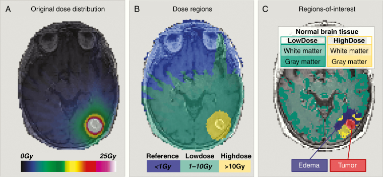
Forty patients with brain metastases from non-small cell lung cancer ( = 26) and malignant melanoma ( = 14) received SRS (15-25 Gy). Longitudinal MRI was performed pre-SRS and at 3, 6, 9, 12, and 18 months post-SRS. Measures of tissue diffusivity and vascularity were assessed by diffusion-weighted and perfusion MRI, respectively. All maps were normalized to white matter receiving less than 1 Gy. Longitudinal responses were assessed in normal-appearing brain, excluding tumor and edema, in the LowDose (1-10 Gy) and HighDose (>10 Gy) regions. The Eastern Cooperative Oncology Group (ECOG) performance status was recorded pre-SRS.

RESULTS

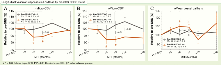
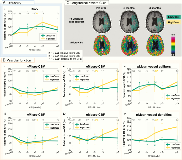
Following SRS, the diffusivity in the LowDose region increased continuously for 1 year (105.1% ± 6.2%; < .001), before reversing toward pre-SRS levels at 18 months. Transient reductions in microvascular cerebral blood volume ( < .05), blood flow ( < .05), and vessel densities ( < .05) were observed in LowDose at 6-9 months post-SRS. Correspondingly, vessel calibers in LowDose transiently increased at 3-9 months ( < .01). The responses in HighDose displayed similar trends as in LowDose, but with larger interpatient variations. Vascular responses followed pre-SRS ECOG status.

CONCLUSIONS

Our results imply that even low doses of radiation to normal-appearing brain following cerebral SRS induce increased diffusivity and reduced vascular function for up until 18 months. In particular, the vascular responses indicate the reduced ability of the normal-appearing brain tissue to form new capillaries. Assessing the potential long-term neurologic effects of SRS on the normal-appearing brain is warranted.

摘要

背景

立体定向放射外科治疗(SRS)脑转移瘤后,MRI 可能有助于深入了解受照射的正常脑组织在扩散率和血管功能方面的纵向变化。

方法

40 例非小细胞肺癌(n = 26)和恶性黑色素瘤(n = 14)脑转移患者接受了 SRS(15 - 25 Gy)。在 SRS 前以及 SRS 后 3、6、9、12 和 18 个月进行纵向 MRI 检查。分别通过扩散加权 MRI 和灌注 MRI 评估组织扩散率和血管情况。所有图像均标准化至接受剂量小于 1 Gy 的白质。在低剂量(1 - 10 Gy)和高剂量(>10 Gy)区域,对排除肿瘤和水肿的正常脑组织进行纵向反应评估。在 SRS 前记录东部肿瘤协作组(ECOG)体能状态。

结果

SRS 后,低剂量区域的扩散率持续增加 1 年(105.1% ± 6.2%;P <.001),在 18 个月时恢复到 SRS 前水平。在 SRS 后 6 - 9 个月,低剂量区域观察到微血管脑血容量(P <.05)、血流(P <.05)和血管密度(P <.05)的短暂降低。相应地,低剂量区域的血管管径在 3 - 9 个月时短暂增加(P <.01)。高剂量区域的反应与低剂量区域相似,但患者间差异更大。血管反应与 SRS 前的 ECOG 状态相关。

结论

我们的结果表明,即使是脑部 SRS 后对正常脑组织的低剂量辐射,在长达 18 个月内也会导致扩散率增加和血管功能降低。特别是,血管反应表明正常脑组织形成新毛细血管的能力降低。有必要评估 SRS 对正常脑组织潜在的长期神经学影响。

https://cdn.ncbi.nlm.nih.gov/pmc/blobs/f8fd/7212876/1eb68d1f9e5f/vdaa028f0004.jpg
https://cdn.ncbi.nlm.nih.gov/pmc/blobs/f8fd/7212876/5b945a3f4474/vdaa028f0001.jpg
https://cdn.ncbi.nlm.nih.gov/pmc/blobs/f8fd/7212876/03e6d30c9b83/vdaa028f0002.jpg
https://cdn.ncbi.nlm.nih.gov/pmc/blobs/f8fd/7212876/94b2fc33c957/vdaa028f0003.jpg
https://cdn.ncbi.nlm.nih.gov/pmc/blobs/f8fd/7212876/1eb68d1f9e5f/vdaa028f0004.jpg

相似文献

1
Responses in the diffusivity and vascular function of the irradiated normal brain are seen up until 18 months following SRS of brain metastases.在脑转移瘤立体定向放射治疗(SRS)后长达18个月的时间里,均可观察到受照射的正常脑组织在扩散率和血管功能方面的变化。
Neurooncol Adv. 2020 Feb 28;2(1):vdaa028. doi: 10.1093/noajnl/vdaa028. eCollection 2020 Jan-Dec.
2
Brain metastases with poor vascular function are susceptible to pseudoprogression after stereotactic radiation surgery.血管功能较差的脑转移瘤在立体定向放射治疗后易发生假性进展。
Adv Radiat Oncol. 2018 May 17;3(4):559-567. doi: 10.1016/j.adro.2018.05.005. eCollection 2018 Oct-Dec.
3
Diffusion and Perfusion MRI Predicts Response Preceding and Shortly After Radiosurgery to Brain Metastases: A Pilot Study.弥散和灌注 MRI 预测脑转移放射外科治疗前后的反应:一项初步研究。
J Neuroimaging. 2021 Mar;31(2):317-323. doi: 10.1111/jon.12828. Epub 2020 Dec 28.
4
Adverse radiation effect after stereotactic radiosurgery for brain metastases: incidence, time course, and risk factors.脑转移瘤立体定向放射治疗后的不良放射效应:发生率、时间进程及危险因素
J Neurosurg. 2015 Aug;123(2):373-86. doi: 10.3171/2014.10.JNS141610. Epub 2015 May 15.
5
Stereotactic radiosurgery for newly diagnosed brain metastases: comparison of three dose levels.立体定向放射外科治疗新诊断的脑转移瘤:三种剂量水平的比较。
Strahlenther Onkol. 2014 Sep;190(9):786-91. doi: 10.1007/s00066-014-0625-1. Epub 2014 Mar 25.
6
Stereotactic radiosurgery for the treatment of melanoma and renal cell carcinoma brain metastases.立体定向放射外科治疗黑色素瘤和肾细胞癌脑转移瘤。
Oncol Rep. 2013 Feb;29(2):407-12. doi: 10.3892/or.2012.2139. Epub 2012 Nov 14.
7
Impact of the radiosurgery prescription dose on the local control of small (2 cm or smaller) brain metastases.伽玛刀处方剂量对 2cm 或以下小(脑)转移瘤局部控制的影响。
J Neurosurg. 2017 Mar;126(3):735-743. doi: 10.3171/2016.3.JNS153014. Epub 2016 May 27.
8
Investigating diffusion patterns of brain metastases pre- and post-stereotactic radiosurgery: a feasibility study.探讨立体定向放射外科前后脑转移瘤扩散模式:一项可行性研究。
Biomed Phys Eng Express. 2021 Sep 3;7(6). doi: 10.1088/2057-1976/ac1d89.
9
Impact of 2-staged stereotactic radiosurgery for treatment of brain metastases ≥ 2 cm.2 期立体定向放射外科治疗 ≥ 2 cm 的脑转移瘤的影响。
J Neurosurg. 2018 Aug;129(2):366-382. doi: 10.3171/2017.3.JNS162532. Epub 2017 Sep 22.
10
MRI Detection of Changes in Tissue Sodium Concentration in Brain Metastases after Stereotactic Radiosurgery: A Feasibility Study.磁共振成像检测立体定向放射外科后脑转移灶组织钠离子浓度变化:一项可行性研究。
J Neuroimaging. 2021 Mar;31(2):297-305. doi: 10.1111/jon.12823. Epub 2020 Dec 22.

引用本文的文献

1
Ionizing radiation, cerebrovascular disease, and consequent dementia: A review and proposed framework relevant to space radiation exposure.电离辐射、脑血管疾病及由此引发的痴呆症:与太空辐射暴露相关的综述及框架建议
Front Physiol. 2022 Oct 25;13:1008640. doi: 10.3389/fphys.2022.1008640. eCollection 2022.
2
Evaluation of normal-appearing white matter with perfusion and diffusion MRI in patients with treated glioblastoma.评估经治疗的胶质母细胞瘤患者的正常表现的白质的灌注和弥散 MRI。
MAGMA. 2022 Feb;35(1):153-162. doi: 10.1007/s10334-021-00990-5. Epub 2021 Dec 24.
3
A systematic review on the use of quantitative imaging to detect cancer therapy adverse effects in normal-appearing brain tissue.

本文引用的文献

1
Identifying early diffusion imaging biomarkers of regional white matter injury as indicators of executive function decline following brain radiotherapy: A prospective clinical trial in primary brain tumor patients.识别早期弥散成像生物标志物,作为脑放疗后执行功能下降的区域白质损伤指标:原发性脑肿瘤患者的前瞻性临床试验。
Radiother Oncol. 2019 Mar;132:27-33. doi: 10.1016/j.radonc.2018.11.018. Epub 2018 Dec 20.
2
Cognitive effects of stereotactic radiosurgery in adult patients with brain metastases: A systematic review.立体定向放射外科治疗对成年脑转移瘤患者的认知影响:一项系统评价。
Adv Radiat Oncol. 2018 Jul 11;3(4):568-581. doi: 10.1016/j.adro.2018.06.003. eCollection 2018 Oct-Dec.
3
系统评价使用定量成像技术检测正常外观脑组织中癌症治疗不良反应的研究。
MAGMA. 2022 Feb;35(1):163-186. doi: 10.1007/s10334-021-00985-2. Epub 2021 Dec 17.
4
MR Image Changes of Normal-Appearing Brain Tissue after Radiotherapy.放疗后正常脑组织的磁共振成像变化
Cancers (Basel). 2021 Mar 29;13(7):1573. doi: 10.3390/cancers13071573.
Brain metastases with poor vascular function are susceptible to pseudoprogression after stereotactic radiation surgery.
血管功能较差的脑转移瘤在立体定向放射治疗后易发生假性进展。
Adv Radiat Oncol. 2018 May 17;3(4):559-567. doi: 10.1016/j.adro.2018.05.005. eCollection 2018 Oct-Dec.
4
Radiation-Induced Endothelial Vascular Injury: A Review of Possible Mechanisms.辐射诱导的内皮血管损伤:可能机制综述
JACC Basic Transl Sci. 2018 Aug 28;3(4):563-572. doi: 10.1016/j.jacbts.2018.01.014. eCollection 2018 Aug.
5
Perfusion Magnetic Resonance Imaging Changes in Normal Appearing Brain Tissue after Radiotherapy in Glioblastoma Patients may Confound Longitudinal Evaluation of Treatment Response.胶质母细胞瘤患者放疗后正常脑组织灌注磁共振成像的变化可能会混淆治疗反应的纵向评估。
Radiol Oncol. 2018 Jun 6;52(2):143-151. doi: 10.2478/raon-2018-0022. eCollection 2018 Jun.
6
Stereotactic Radiosurgery in the Management of Patients With Brain Metastases of Non-Small Cell Lung Cancer: Indications, Decision Tools and Future Directions.立体定向放射外科治疗非小细胞肺癌脑转移患者:适应证、决策工具及未来方向
Front Oncol. 2018 May 9;8:154. doi: 10.3389/fonc.2018.00154. eCollection 2018.
7
Functional vascular contributions to cognitive impairment and dementia: mechanisms and consequences of cerebral autoregulatory dysfunction, endothelial impairment, and neurovascular uncoupling in aging.功能血管对认知障碍和痴呆的影响:衰老过程中脑自动调节功能障碍、内皮功能损伤及神经血管解偶联的机制与后果
Am J Physiol Heart Circ Physiol. 2017 Jan 1;312(1):H1-H20. doi: 10.1152/ajpheart.00581.2016. Epub 2016 Oct 28.
8
Early and late effects of radiochemotherapy on cerebral blood flow in glioblastoma patients measured with non-invasive perfusion MRI.采用非侵入性灌注磁共振成像测量胶质母细胞瘤患者放化疗对脑血流量的早期和晚期影响。
Radiother Oncol. 2016 Jan;118(1):24-8. doi: 10.1016/j.radonc.2015.12.017. Epub 2015 Dec 30.
9
Non Tumor Perfusion Changes Following Stereotactic Radiosurgery to Brain Metastases.脑转移瘤立体定向放射治疗后的非肿瘤灌注变化
Technol Cancer Res Treat. 2015 Aug;14(4):497-503. doi: 10.1177/1533034614600279.
10
Vessel caliber--a potential MRI biomarker of tumour response in clinical trials.血管管径——临床试验中肿瘤反应的一种潜在磁共振成像生物标志物。
Nat Rev Clin Oncol. 2014 Oct;11(10):566-84. doi: 10.1038/nrclinonc.2014.126. Epub 2014 Aug 12.