Department of Diagnostic and Interventional Neuroradiology, Medical School Munich, Klinikum Rechts der Isar, Technische Universität München, Ismaninger Str. 22, 81765, Munich, Germany.
Department of Oral and Maxillofacial Surgery and Facial Plastic Surgery, Klinikum Rechts Der Isar, Technische Universität München, Munich, Germany.
Sci Rep. 2020 Jul 14;10(1):11566. doi: 10.1038/s41598-020-68501-5.
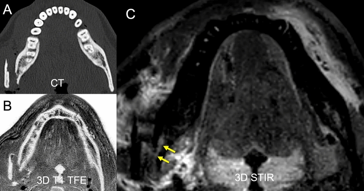
The purpose of this study was to evaluate a magnetic resonance imaging (MRI) protocol for direct visualization of the inferior alveolar nerve in the setting of mandibular fractures. Fifteen patients suffering from unilateral mandible fractures involving the inferior alveolar nerve (15 affected IAN and 15 unaffected IAN from contralateral side) were examined on a 3 T scanner (Elition, Philips Healthcare, Best, the Netherlands) and compared with 15 healthy volunteers (30 IAN in total). The sequence protocol consisted of a 3D STIR, 3D DESS and 3D T1 FFE sequence. Apparent nerve-muscle contrast-to-noise ratio (aNMCNR), apparent signal-to-noise ratio (aSNR), nerve diameter and fracture dislocation were evaluated by two radiologists and correlated with nerve impairment. Furthermore, dislocation as depicted by MRI was compared to computed tomography (CT) images. Patients with clinically evident nerve impairment showed a significant increase of aNMCNR, aSNR and nerve diameter compared to healthy controls and to the contralateral side (p < 0.05). Furthermore, the T1 FFE sequence allowed dislocation depiction comparable to CT. This prospective study provides a rapid imaging protocol using the 3D STIR and 3D T1 FFE sequence that can directly assess both mandible fractures and IAN damage. In patients with hypoesthesia following mandibular fractures, increased aNMCNR, aSNR and nerve diameter on MRI imaging may help identify patients with a risk of prolonged or permanent hypoesthesia at an early time.
本研究旨在评估一种磁共振成像 (MRI) 方案,用于直接观察下颌骨骨折中下颌下神经的情况。 15 名单侧下颌骨骨折伴有下颌下神经损伤的患者(15 条患侧 IAN 和 15 条对侧未受损 IAN)在 3T 扫描仪(荷兰飞利浦医疗公司的 Elition)上进行了检查,并与 15 名健康志愿者(共 30 条 IAN)进行了比较。序列方案包括 3D STIR、3D DESS 和 3D T1 FFE 序列。两位放射科医生评估了表观神经-肌肉对比噪声比(aNMCNR)、表观信噪比(aSNR)、神经直径和骨折脱位,并与神经损伤相关联。此外,还将 MRI 显示的脱位与 CT 图像进行了比较。有临床明显神经损伤的患者的 aNMCNR、aSNR 和神经直径与健康对照组和对侧相比显著增加(p<0.05)。此外,T1 FFE 序列可以与 CT 一样清晰地显示脱位。这项前瞻性研究提供了一种使用 3D STIR 和 3D T1 FFE 序列的快速成像方案,可直接评估下颌骨骨折和 IAN 损伤。在下颌骨骨折后出现感觉减退的患者中,MRI 成像上的 aNMCNR、aSNR 和神经直径增加可能有助于早期识别有长期或永久性感觉减退风险的患者。