Ülker Ersin, Kirtiloğlu Tuğrul, Taban Burcu
Research Assistant, Department of Periodontology, Faculty of Dentistry, Ondokuz Mayis University, Samsun, Turkey.
Associated Proffessor, Department of Periodontology, Faculty of Dentistry, Ondokuz Mayis University, Samsun, Turkey.
J Clin Exp Dent. 2020 Jun 1;12(6):e607-e609. doi: 10.4317/jced.56757. eCollection 2020 Jun.
Ameloblastoma is a rare tumor which develops from odontogenic epithelium and its remnants and it occurs in the jaws. Peripheral ameloblastomas are rare and benign extraooseous ameloblastomas which effects soft tissues. This case report declares a peripheral ameloblastoma which is a rare type of ameloblastoma.
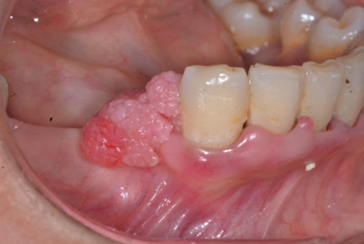
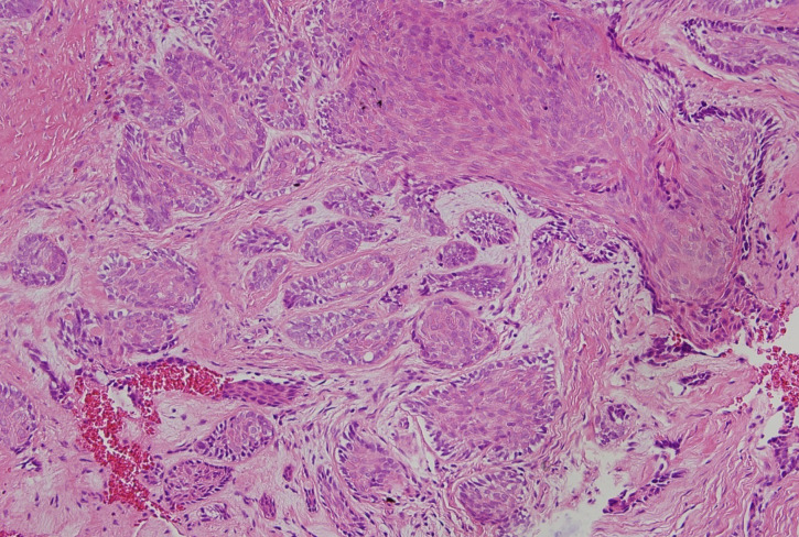
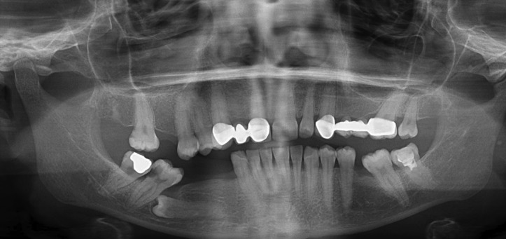
34 year old female patient referred with a complaint of a gingival growth at right lower premolar area. A firm and granular surfaced gingival growth with the color of pink and red and having 1.5x1 cm sizes was observed at the mentioned area. With an incision from lower right second incisor tooth to lower right second molar tooth a flap from bone was made and lesion was excised. After then specimen was submitted to histopathologic examination. After clinical, radiological and pathological examinations lesion was described as peripheral ameloblastoma.
At the control examination after three months of excision there was no recurrence and patieant has no complaint.
Although reccurens rate of peripheral ameloblastomas are low, long-term follow-ups are suggested Patient was informed about the importance of regular controls for early diagnosis of possible reccurenses and regular controls were made during one year after excision. Peripheral ameloblastoma, gingiva, gingival hyperplasia, gingival lesion, alveolar mucosa, extraosseous.
成釉细胞瘤是一种罕见的肿瘤,由牙源性上皮及其残余组织发展而来,发生于颌骨。外周型成釉细胞瘤是罕见的良性骨外型成釉细胞瘤,累及软组织。本病例报告的是一例外周型成釉细胞瘤,这是一种罕见类型的成釉细胞瘤。
一名34岁女性患者因右下前磨牙区牙龈肿物前来就诊。在上述区域观察到一个质地坚实、表面呈颗粒状的牙龈肿物,颜色为粉红色和红色,大小为1.5×1厘米。从右下第二切牙至右下第二磨牙做切口,掀起骨膜瓣,切除病变组织。随后将标本送检进行组织病理学检查。经临床、影像学和病理学检查,病变被诊断为外周型成釉细胞瘤。
切除术后三个月的复查中未见复发,患者无不适主诉。
尽管外周型成釉细胞瘤的复发率较低,但仍建议进行长期随访。已告知患者定期复查对于早期诊断可能复发的重要性,并在切除术后一年内进行了定期复查。外周型成釉细胞瘤、牙龈、牙龈增生、牙龈病变、牙槽黏膜、骨外型