NIHR Leeds Biomedical Research Centre, Leeds Teaching Hospitals NHS Trust, Leeds, UK.
Medical Physics and Engineering, Leeds Teaching Hospitals NHS Trust, Leeds, UK.
Eur Radiol. 2020 Dec;30(12):6603-6613. doi: 10.1007/s00330-020-06999-z. Epub 2020 Jul 15.
To assess the ability of quantitative T2, diffusion tensor imaging (DTI) and radiologist's scores to detect muscle changes following acute muscle tear in soccer and rugby players. To assess the ability of these parameters to predict return to play times.
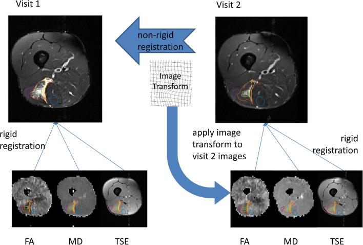
In this prospective, longitudinal study, 13 male athletes (age 19 to 34 years; mean 25 years) underwent MRI within 1 week of suffering acute muscle tear. Imaging included measurements of T2 and DTI parameters. Images were also assessed using modified Peetrons and British athletics muscle injury classification (BAMIC) scores. Participants returned for a second scan within 1 week of being determined fit to return to play. MRI measurements were compared between visits. Pearson's correlation between visit 1 measurements and return to play times was assessed.
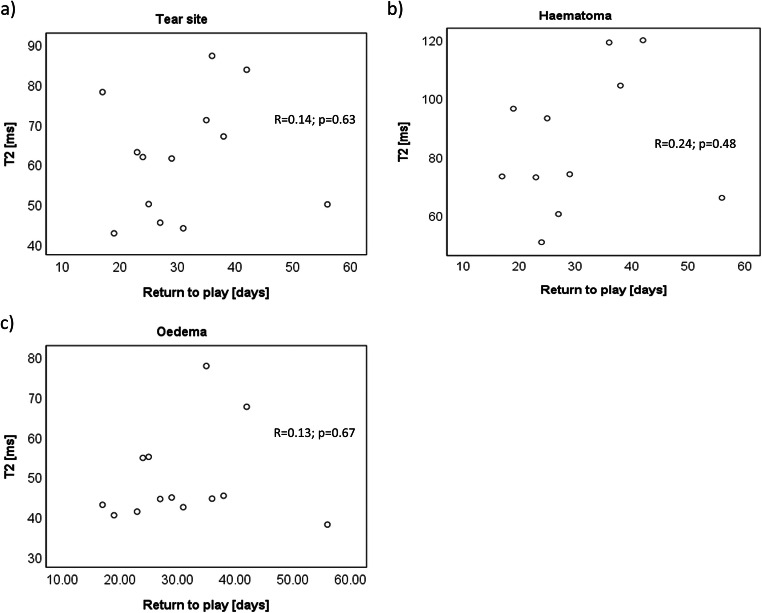
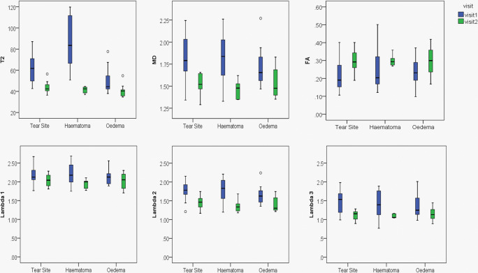
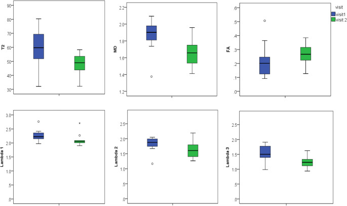
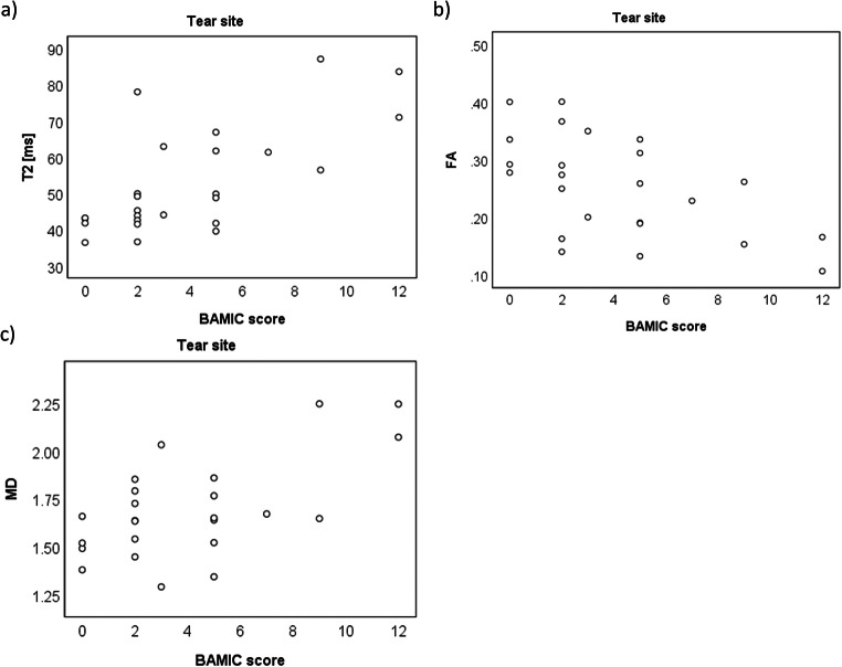
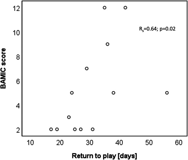
There were significant differences between visits in BAMIC scores (Z = - 2.088; p = 0.037), modified Peetrons (Z = - 2.530; p = 0.011) and quantitative MRI measurements; T2, 13.12 ms (95% CI, 4.82 ms, 21.42 ms; p = 0.01); mean diffusivity (0.22 (0.04, 0.39); p = 0.02) and fractional anisotropy (0.07 (0.01, 0.14); p = 0.03). BAMIC scores showed a significant correlation with return to play time (R = 0.64; p = 0.02), but modified Peetrons scores and quantitative parameters did not.
T2 and DTI measurements in muscle can detect changes due to healing following muscle tear. Although BAMIC scores correlated well with return to play times, in this small study, quantitative MRI values did not, suggesting that T2 and DTI measurements are inferior predictors of return to play time compared with visual scoring.
• Muscle changes following acute muscle tear can be measured using T2 and diffusion measurements on MRI. • Measurements of T2 and diffusion using MRI are not as good as a radiologist's visual report at predicting return to play time after acute muscle tear.
评估定量 T2、弥散张量成像(DTI)和放射科医生评分在检测足球和橄榄球运动员急性肌肉撕裂后肌肉变化方面的能力。评估这些参数预测重返赛场时间的能力。
在这项前瞻性、纵向研究中,13 名男性运动员(年龄 19 至 34 岁;平均 25 岁)在急性肌肉撕裂后 1 周内接受 MRI 检查。成像包括 T2 和 DTI 参数的测量。还使用改良 Peetrons 和英国田径肌肉损伤分类(BAMIC)评分评估图像。参与者在确定适合重返赛场后 1 周内进行第二次扫描。比较两次就诊时的 MRI 测量值。评估第 1 次就诊时测量值与重返赛场时间之间的 Pearson 相关性。
BAMIC 评分(Z=-2.088;p=0.037)、改良 Peetrons 评分(Z=-2.530;p=0.011)和定量 MRI 测量值在就诊时存在显著差异;T2,13.12ms(95%CI,4.82ms,21.42ms;p=0.01);平均弥散系数(0.22(0.04,0.39);p=0.02)和各向异性分数(0.07(0.01,0.14);p=0.03)。BAMIC 评分与重返赛场时间呈显著相关性(R=0.64;p=0.02),但改良 Peetrons 评分和定量参数没有相关性。
肌肉中的 T2 和 DTI 测量值可以检测肌肉撕裂愈合后的变化。尽管 BAMIC 评分与重返赛场时间相关性良好,但在这项小型研究中,定量 MRI 值没有相关性,这表明 T2 和 DTI 测量值与视觉评分相比,预测急性肌肉撕裂后重返赛场时间的效果较差。
• MRI 上的 T2 和弥散测量可用于测量急性肌肉撕裂后的肌肉变化。• MRI 上 T2 和弥散的测量值不如放射科医生的视觉报告,在预测急性肌肉撕裂后重返赛场时间方面效果较差。