Gough Sarah L, Anderson Jonathan D C, Dixon Jonathon J
Rainbow Equine Hospital, North Yorkshire, United Kingdom.
J Vet Intern Med. 2020 Sep;34(5):2142-2151. doi: 10.1111/jvim.15848. Epub 2020 Jul 24.
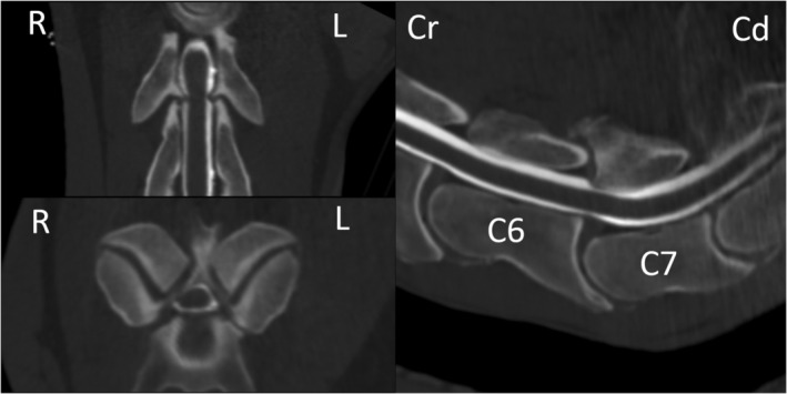
Three-dimensional computed tomographic (CT) evaluation of the cervical vertebral column enables more accurate identification of osseous and soft tissue lesions than traditional latero-lateral radiography. However, examination of the complete cervical vertebral column has been limited by horse size, preventing evaluation of the caudal cervical vertebrae.
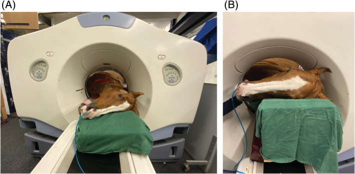
To describe a technique to enable CT myelography of the complete cervical spine and describe the findings in 51 horses.
Records of 51 horses presented for evaluation of cervical vertebral lesions.
A retrospective review of clinical records from all horses presented for CT myelography to further investigate possible cervical vertebral lesions was performed. A description of a novel approach to CT myelography in horses and retrospective review of the findings in clinical cases has been included.
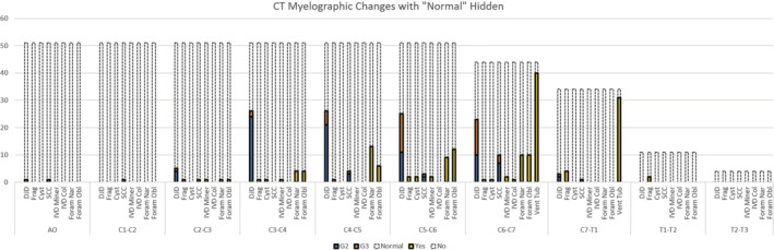
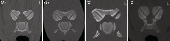
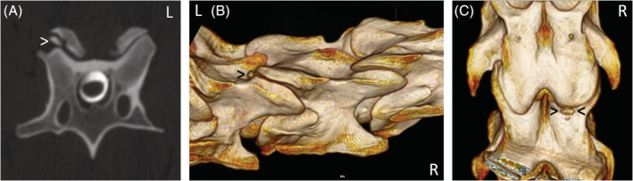
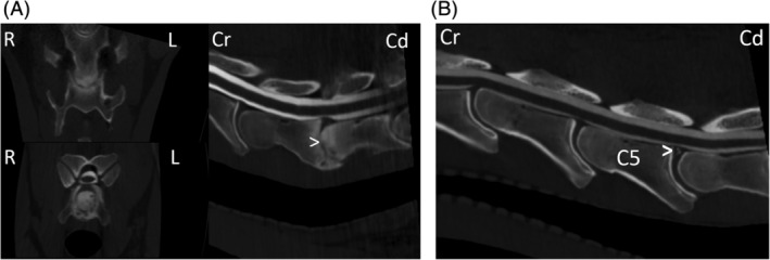
Degenerative joint disease was identified at 1 or more dorsal articular process joint in 50/51 horses, of which 44/51 had a site of grade 2 or greater. Spinal cord compression was observed on CT myelography in 31/51 horses, whereas attenuation of the dorsal contrast column was identified radiographically in 11/50 horses. Thirty-three horses showed narrowing or obliteration of the intervertebral foramina at 1 or more site and osteochondral fragments were seen in 11/51 horses.
Computed tomography myelography is relatively safe and an easily performed technique with the correct equipment, enabling evaluation of the cervical vertebral structures of horses in all planes and volumetrically. It is possible that lesion extent might be underestimated with this diagnostic modality, hence interpretation should be complimented with flexed and extended views radiographically.
与传统的侧位X线摄影相比,颈椎的三维计算机断层扫描(CT)评估能够更准确地识别骨和软组织病变。然而,完整颈椎的检查受到马匹体型的限制,无法对颈椎尾段进行评估。
描述一种用于对整个颈椎进行CT脊髓造影的技术,并描述51匹马的检查结果。
51匹因颈椎病变接受评估的马匹记录。
对所有接受CT脊髓造影以进一步调查可能的颈椎病变的马匹的临床记录进行回顾性分析。其中包括对一种用于马匹CT脊髓造影的新方法的描述以及对临床病例检查结果的回顾性分析。
51匹马中有50匹在1个或多个背侧关节突关节处发现退行性关节病,其中51匹马中有44匹存在2级或更高级别的病变部位。51匹马中有31匹在CT脊髓造影中观察到脊髓受压,而50匹马中有11匹在X线片上发现背侧造影剂柱衰减。33匹马在1个或多个部位出现椎间孔狭窄或闭塞,51匹马中有11匹可见骨软骨碎片。
计算机断层扫描脊髓造影相对安全,使用正确的设备易于操作,能够在所有平面和容积上评估马的颈椎结构。这种诊断方式可能会低估病变范围,因此在解读时应辅以X线片的屈曲和伸展位视图。