• 文献检索
  • 文档翻译
  • 深度研究
  • 学术资讯
  • Suppr Zotero 插件Zotero 插件
  • 邀请有礼
  • 套餐&价格
  • 历史记录
应用&插件
Suppr Zotero 插件Zotero 插件浏览器插件Mac 客户端Windows 客户端微信小程序
定价
高级版会员购买积分包购买API积分包
服务
文献检索文档翻译深度研究API 文档MCP 服务
关于我们
关于 Suppr公司介绍联系我们用户协议隐私条款
关注我们

Suppr 超能文献

核心技术专利:CN118964589B侵权必究
粤ICP备2023148730 号-1Suppr @ 2026

文献检索

告别复杂PubMed语法,用中文像聊天一样搜索,搜遍4000万医学文献。AI智能推荐,让科研检索更轻松。

立即免费搜索

文件翻译

保留排版,准确专业,支持PDF/Word/PPT等文件格式,支持 12+语言互译。

免费翻译文档

深度研究

AI帮你快速写综述,25分钟生成高质量综述,智能提取关键信息,辅助科研写作。

立即免费体验

新型鼠类脑海绵状血管畸形模型。

Novel Murine Models of Cerebral Cavernous Malformations.

机构信息

Department of Molecular Genetics and Microbiology, Duke University School of Medicine, Durham, NC, 27705, USA.

Neurovascular Surgery Program, Department of Neurosurgery, University of Chicago Medicine and Biological Sciences, Chicago, IL, 60637, USA.

出版信息

Angiogenesis. 2020 Nov;23(4):651-666. doi: 10.1007/s10456-020-09736-8. Epub 2020 Jul 24.

DOI:10.1007/s10456-020-09736-8
PMID:32710309
原文链接:https://pmc.ncbi.nlm.nih.gov/articles/PMC7530029/
Abstract

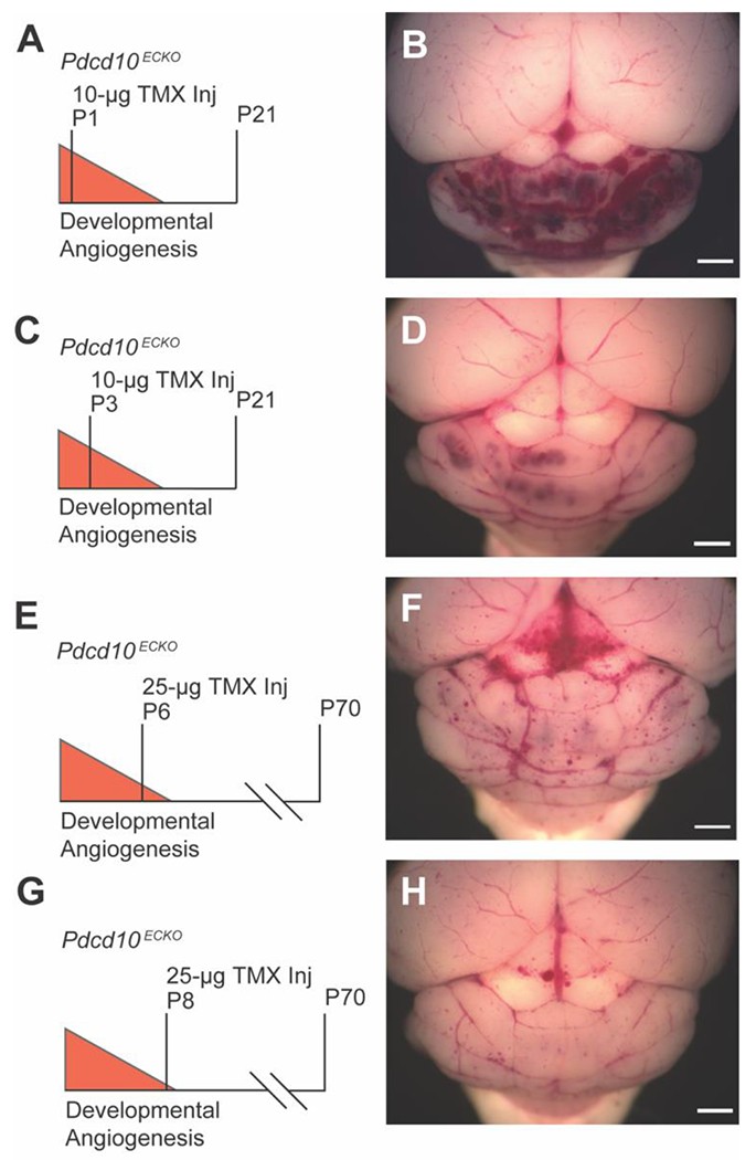
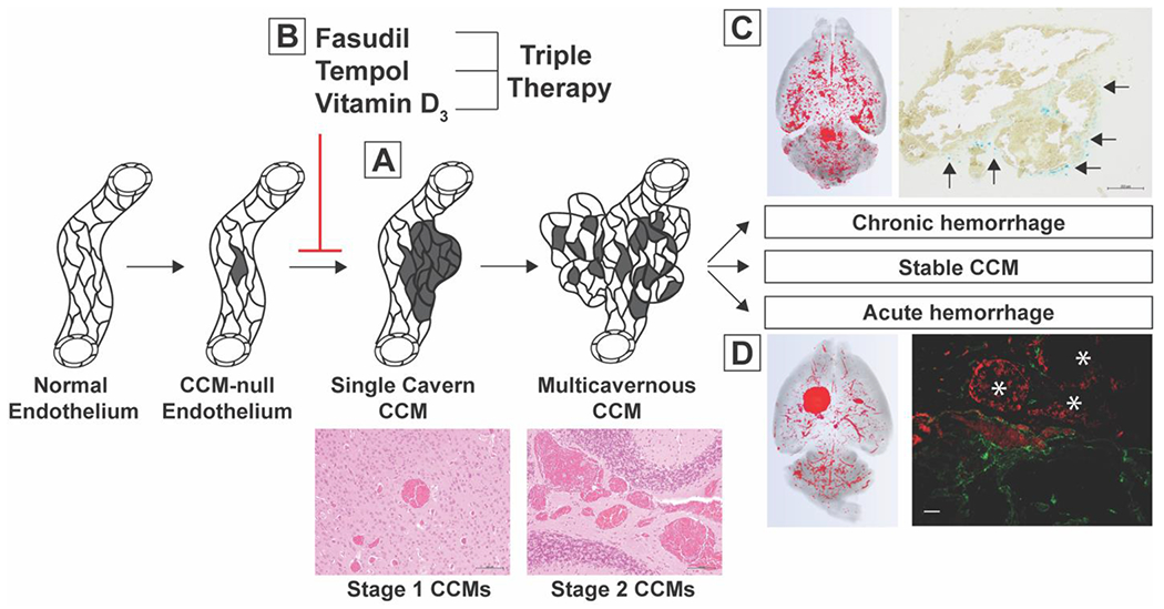
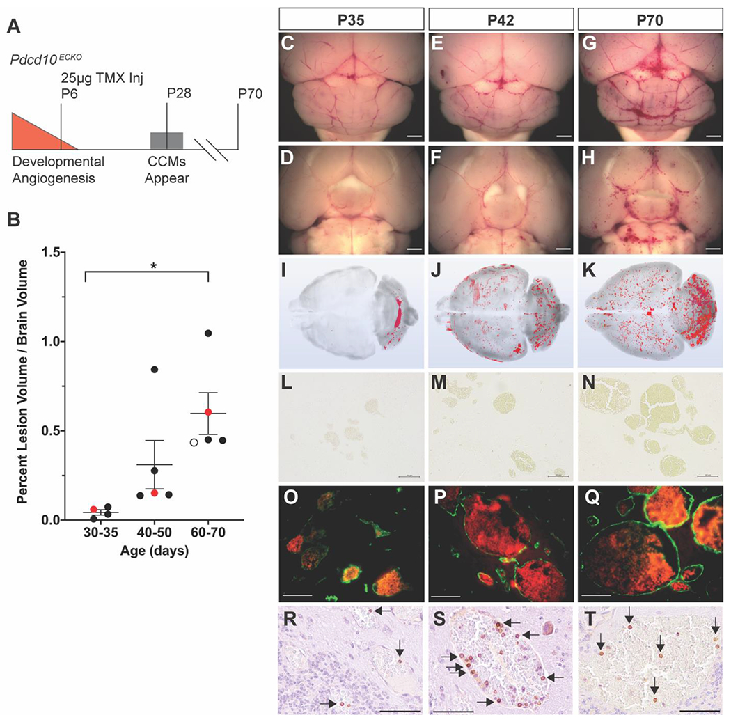
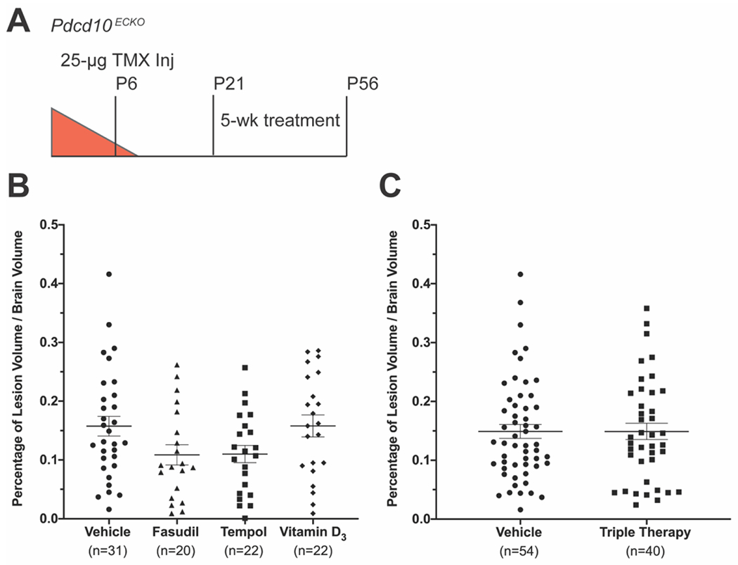
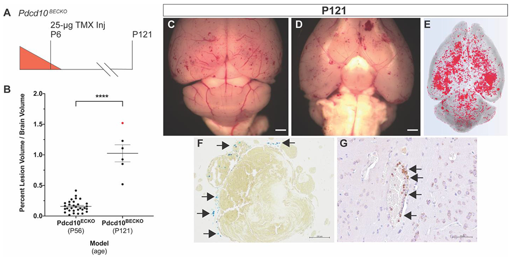
Cerebral cavernous malformations (CCMs) are ectatic capillary-venous malformations that develop in approximately 0.5% of the population. Patients with CCMs may develop headaches, focal neurologic deficits, seizures, and hemorrhages. While symptomatic CCMs, depending upon the anatomic location, can be surgically removed, there is currently no pharmaceutical therapy to treat CCMs. Several mouse models have been developed to better understand CCM pathogenesis and test therapeutics. The most common mouse models induce a large CCM burden that is anatomically restricted to the cerebellum and contributes to lethality in the early days of life. These inducible models thus have a relatively short period for drug administration. We developed an inducible CCM3 mouse model that develops CCMs after weaning and provides a longer period for potential therapeutic intervention. Using this new model, three recently proposed CCM therapies, fasudil, tempol, vitamin D, and a combination of the three drugs, failed to substantially reduce CCM formation when treatment was administered for 5 weeks, from postnatal day 21 (P21) to P56. We next restricted Ccm3 deletion to the brain vasculature and provided greater time (121 days) for CCMs to develop chronic hemorrhage, recapitulating the human lesions. We also developed the first model of acute CCM hemorrhage by injecting mice harboring CCMs with lipopolysaccharide. These efficient models will enable future drug studies to more precisely target clinically relevant features of CCM disease: CCM formation, chronic hemorrhage, and acute hemorrhage.

摘要

脑内海绵状血管畸形(CCMs)是一种扩张的毛细血管-静脉畸形,约占人口的 0.5%。CCMs 患者可能会出现头痛、局灶性神经功能缺损、癫痫发作和出血。虽然有症状的 CCMs 根据解剖位置可以通过手术切除,但目前尚无药物治疗方法。已经开发了几种小鼠模型来更好地了解 CCM 发病机制并测试治疗方法。最常见的小鼠模型诱导大量 CCM 负担,解剖上局限于小脑,并导致生命早期的致死性。因此,这些诱导型模型用于药物给药的时间相对较短。我们开发了一种诱导型 CCM3 小鼠模型,该模型在断奶后会发展出 CCMs,并为潜在的治疗干预提供了更长的时间。使用这种新模型,三种最近提出的 CCM 治疗方法(法舒地尔、替米沙坦、维生素 D 以及三种药物的联合应用)在从出生后第 21 天(P21)至 P56 的 5 周治疗期间未能显著减少 CCM 的形成。接下来,我们将 Ccm3 缺失限制在脑血管中,并提供更长的时间(121 天)让 CCMs 发展慢性出血,重现人类病变。我们还通过向携带 CCM 的小鼠注射脂多糖来开发急性 CCM 出血的首个模型。这些高效模型将使未来的药物研究能够更精确地针对 CCM 疾病的临床相关特征:CCM 的形成、慢性出血和急性出血。

https://cdn.ncbi.nlm.nih.gov/pmc/blobs/2e06/7530029/92d88a930431/nihms-1614973-f0006.jpg
https://cdn.ncbi.nlm.nih.gov/pmc/blobs/2e06/7530029/2185bc7f6d7f/nihms-1614973-f0001.jpg
https://cdn.ncbi.nlm.nih.gov/pmc/blobs/2e06/7530029/fb4375555838/nihms-1614973-f0002.jpg
https://cdn.ncbi.nlm.nih.gov/pmc/blobs/2e06/7530029/2f0879260143/nihms-1614973-f0003.jpg
https://cdn.ncbi.nlm.nih.gov/pmc/blobs/2e06/7530029/a93f21b8ca58/nihms-1614973-f0004.jpg
https://cdn.ncbi.nlm.nih.gov/pmc/blobs/2e06/7530029/b1a075921a81/nihms-1614973-f0005.jpg
https://cdn.ncbi.nlm.nih.gov/pmc/blobs/2e06/7530029/92d88a930431/nihms-1614973-f0006.jpg

相似文献

1
Novel Murine Models of Cerebral Cavernous Malformations.新型鼠类脑海绵状血管畸形模型。
Angiogenesis. 2020 Nov;23(4):651-666. doi: 10.1007/s10456-020-09736-8. Epub 2020 Jul 24.
2
Mural Cell-Specific Deletion of Cerebral Cavernous Malformation 3 in the Brain Induces Cerebral Cavernous Malformations.脑内血管性腔隙畸形 3 的壁细胞特异性缺失可诱导脑内血管性腔隙畸形。
Arterioscler Thromb Vasc Biol. 2020 Sep;40(9):2171-2186. doi: 10.1161/ATVBAHA.120.314586. Epub 2020 Jul 9.
3
Differential angiogenesis function of CCM2 and CCM3 in cerebral cavernous malformations.CCM2 和 CCM3 在脑动静脉畸形中的差异血管生成功能。
Neurosurg Focus. 2010 Sep;29(3):E1. doi: 10.3171/2010.5.FOCUS1090.
4
RhoA Kinase Inhibition With Fasudil Versus Simvastatin in Murine Models of Cerebral Cavernous Malformations.在脑海绵状血管畸形小鼠模型中,法舒地尔与辛伐他汀对RhoA激酶的抑制作用比较
Stroke. 2017 Jan;48(1):187-194. doi: 10.1161/STROKEAHA.116.015013. Epub 2016 Nov 22.
5
Conditional deletion of Ccm2 causes hemorrhage in the adult brain: a mouse model of human cerebral cavernous malformations.条件性敲除 Ccm2 可导致成年大脑出血:人类脑海绵状血管畸形的小鼠模型。
Hum Mol Genet. 2011 Aug 15;20(16):3198-206. doi: 10.1093/hmg/ddr225. Epub 2011 May 19.
6
Familial Cerebral Cavernous Malformations家族性脑海绵状血管畸形
7
Novel Chronic Mouse Model of Cerebral Cavernous Malformations.新型慢性脑动静脉畸形小鼠模型
Stroke. 2020 Apr;51(4):1272-1278. doi: 10.1161/STROKEAHA.119.027207. Epub 2020 Jan 29.
8
mTORC1 Signaling in Brain Endothelial Progenitors Contributes to CCM Pathogenesis.mTORC1 信号在脑内皮祖细胞中的作用促进 CCM 发病机制。
Circ Res. 2024 Aug 2;135(4):e94-e113. doi: 10.1161/CIRCRESAHA.123.324015. Epub 2024 Jul 3.
9
Cerebral Cavernous Malformations Develop Through Clonal Expansion of Mutant Endothelial Cells.脑内海绵状血管畸形通过突变内皮细胞的克隆扩张发展。
Circ Res. 2018 Oct 26;123(10):1143-1151. doi: 10.1161/CIRCRESAHA.118.313970.
10
Caveolae-mediated Tie2 signaling contributes to CCM pathogenesis in a brain endothelial cell-specific Pdcd10-deficient mouse model.小窝介导的 Tie2 信号通路促进了脑内皮细胞特异性 Pdcd10 缺失小鼠模型中 CCM 的发病机制。
Nat Commun. 2021 Jan 25;12(1):504. doi: 10.1038/s41467-020-20774-0.

引用本文的文献

1
Focused ultrasound augments the delivery and penetration of model therapeutics into cerebral cavernous malformations.聚焦超声增强了模型治疗药物向脑海绵状血管畸形的递送和渗透。
J Control Release. 2025 Jul 10;383:113861. doi: 10.1016/j.jconrel.2025.113861. Epub 2025 May 16.
2
Cerebral Cavernous Malformation: From Genetics to Pharmacotherapy.脑海绵状血管畸形:从遗传学至药物治疗
Brain Behav. 2025 Jan;15(1):e70223. doi: 10.1002/brb3.70223.
3
Intravital Imaging of Disease Mechanisms in a Mouse Model of CCM Skin Lesions-Brief Report.

本文引用的文献

1
Novel Chronic Mouse Model of Cerebral Cavernous Malformations.新型慢性脑动静脉畸形小鼠模型
Stroke. 2020 Apr;51(4):1272-1278. doi: 10.1161/STROKEAHA.119.027207. Epub 2020 Jan 29.
2
Symptomatic Brain Hemorrhages from Cavernous Angioma After Botulinum Toxin Injections, a Role of TLR/MEKK3 Mechanism? Case Report and Review of the Literature.肉毒杆菌毒素注射后海绵状血管瘤所致症状性脑内出血,TLR/MEKK3机制的作用?病例报告及文献复习
World Neurosurg. 2020 Apr;136:7-11. doi: 10.1016/j.wneu.2019.12.172. Epub 2020 Jan 7.
3
Distinct cellular roles for PDCD10 define a gut-brain axis in cerebral cavernous malformation.
CCM皮肤病变小鼠模型中疾病机制的活体成像——简报
Arterioscler Thromb Vasc Biol. 2025 Jan;45(1):113-118. doi: 10.1161/ATVBAHA.124.321056. Epub 2024 Nov 21.
4
Force-mediated recruitment and reprogramming of healthy endothelial cells drive vascular lesion growth.力介导的健康内皮细胞募集和重编程驱动血管病变生长。
Nat Commun. 2024 Oct 6;15(1):8660. doi: 10.1038/s41467-024-52866-6.
5
Focused Ultrasound Augments the Delivery and Penetration of Model Therapeutics into Cerebral Cavernous Malformations.聚焦超声增强模型治疗药物向脑海绵状血管畸形的递送与穿透。
bioRxiv. 2025 Jan 23:2024.08.27.609060. doi: 10.1101/2024.08.27.609060.
6
Except for Robust Outliers, Rapamycin Increases Lesion Burden in a Murine Model of Cerebral Cavernous Malformations.除了对强抗值外,雷帕霉素会增加小鼠脑海绵状血管畸形模型中的病灶负担。
Transl Stroke Res. 2025 Jun;16(3):859-867. doi: 10.1007/s12975-024-01270-9. Epub 2024 Jul 9.
7
Generation of -tdTomato Knock-in Mice for Specific Cerebrovascular Endothelial Cell Targeting.用于特定脑血管内皮细胞靶向的 -tdTomato 敲入小鼠的生成。
Int J Mol Sci. 2024 Apr 25;25(9):4666. doi: 10.3390/ijms25094666.
8
Mild Hypoxia Accelerates Cerebral Cavernous Malformation Disease Through CX3CR1-CX3CL1 Signaling.轻度缺氧通过CX3CR1-CX3CL1信号通路加速脑海绵状血管畸形疾病进程。
Arterioscler Thromb Vasc Biol. 2024 Jun;44(6):1246-1264. doi: 10.1161/ATVBAHA.123.320367. Epub 2024 Apr 25.
9
Transcriptomic signatures of individual cell types in cerebral cavernous malformation.脑动静脉畸形中单个细胞类型的转录组特征。
Cell Commun Signal. 2024 Jan 9;22(1):23. doi: 10.1186/s12964-023-01301-2.
10
Magnetic Resonance Imaging of Mouse Cerebral Cavernomas Reveal Differential Lesion Progression and Variable Permeability to Gadolinium.磁共振成像显示小鼠脑内海绵状血管瘤的病变进展存在差异,钆对比剂渗透性也存在差异。
Arterioscler Thromb Vasc Biol. 2023 Jun;43(6):958-970. doi: 10.1161/ATVBAHA.122.318938. Epub 2023 Apr 20.
PDCD10 在不同细胞中的作用定义了脑动静脉畸形的肠脑轴。
Sci Transl Med. 2019 Nov 27;11(520). doi: 10.1126/scitranslmed.aaw3521.
4
VEGF signalling enhances lesion burden in KRIT1 deficient mice.VEGF 信号增强 KRIT1 缺陷型小鼠的病变负担。
J Cell Mol Med. 2020 Jan;24(1):632-639. doi: 10.1111/jcmm.14773. Epub 2019 Nov 20.
5
A Brain-Targeted Orally Available ROCK2 Inhibitor Benefits Mild and Aggressive Cavernous Angioma Disease.一种脑靶向、可口服的 ROCK2 抑制剂可改善轻度和侵袭性海绵状血管畸形疾病。
Transl Stroke Res. 2020 Jun;11(3):365-376. doi: 10.1007/s12975-019-00725-8. Epub 2019 Aug 24.
6
A conserved CCM complex promotes apoptosis non-autonomously by regulating zinc homeostasis.一个保守的 CCM 复合物通过调节锌稳态非自主地促进细胞凋亡。
Nat Commun. 2019 Apr 17;10(1):1791. doi: 10.1038/s41467-019-09829-z.
7
Rho Kinase Inhibition Blunts Lesion Development and Hemorrhage in Murine Models of Aggressive Pdcd10/Ccm3 Disease.Rho 激酶抑制减轻了小鼠侵袭性 Pdcd10/Ccm3 疾病模型中的病变发展和出血。
Stroke. 2019 Mar;50(3):738-744. doi: 10.1161/STROKEAHA.118.024058.
8
Cerebral cavernous malformations form an anticoagulant vascular domain in humans and mice.脑内海绵状血管畸形在人类和小鼠中形成一个抗凝血的血管区域。
Blood. 2019 Jan 17;133(3):193-204. doi: 10.1182/blood-2018-06-856062. Epub 2018 Nov 15.
9
Ponatinib (AP24534) inhibits MEKK3-KLF signaling and prevents formation and progression of cerebral cavernous malformations.泊那替尼(AP24534)抑制 MEKK3-KLF 信号通路,防止脑动静脉畸形的形成和进展。
Sci Adv. 2018 Nov 7;4(11):eaau0731. doi: 10.1126/sciadv.aau0731. eCollection 2018 Nov.
10
The cerebral cavernous malformation disease causing gene KRIT1 participates in intestinal epithelial barrier maintenance and regulation.导致脑动静脉畸形的 KRIT1 基因参与肠道上皮屏障的维持和调节。
FASEB J. 2019 Feb;33(2):2132-2143. doi: 10.1096/fj.201800343R. Epub 2018 Sep 25.