From the Molecular Genetics and Microbiology Department (M.R.D., D.A.S., D.A.M.), Duke University School of Medicine, Durham, NC.
Medical Scientist Training Program (M.R.D.), Duke University School of Medicine, Durham, NC.
Circ Res. 2018 Oct 26;123(10):1143-1151. doi: 10.1161/CIRCRESAHA.118.313970.
Vascular malformations arise in vessels throughout the entire body. Causative genetic mutations have been identified for many of these diseases; however, little is known about the mutant cell lineage within these malformations.
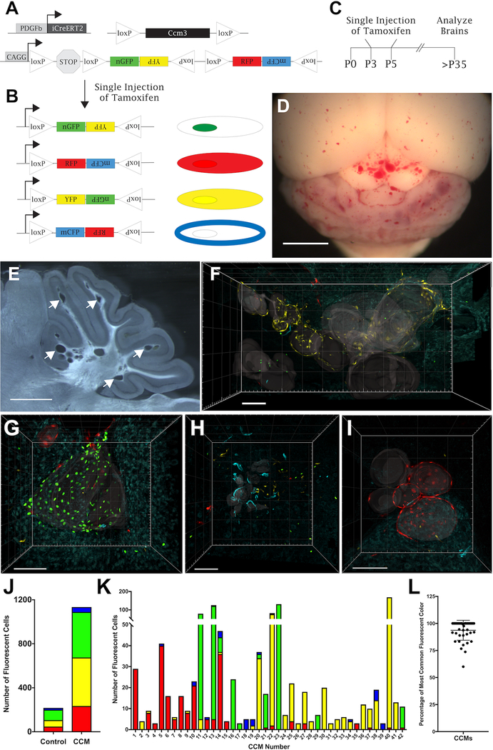
We utilize an inducible mouse model of cerebral cavernous malformations (CCMs) coupled with a multicolor fluorescent reporter to visualize the contribution of mutant endothelial cells (ECs) to the malformation.
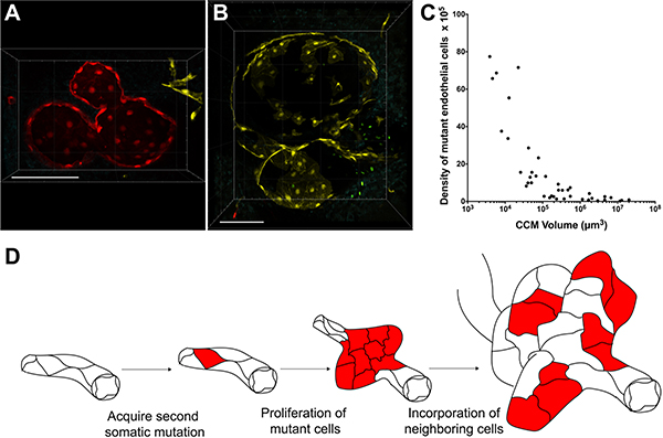
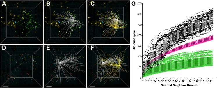
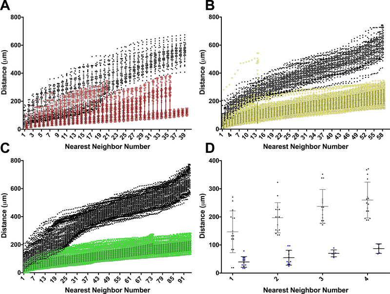
We combined a Ccm3 mouse model with the confetti fluorescent reporter to simultaneously delete Ccm3 and label the mutant EC with 1 of 4 possible colors. We acquired Z-series confocal images from serial brain sections and created 3-dimensional reconstructions of entire CCMs to visualize mutant ECs during CCM development. We observed a pronounced pattern of CCMs lined with mutant ECs labeled with a single confetti color (n=42). The close 3-dimensional distribution, as determined by the nearest neighbor analysis, of the clonally dominant ECs within the CCM was statistically different than the background confetti labeling of ECs in non-CCM control brain slices as well as a computer simulation ( P<0.001). Many of the small (<100 μm diameter) CCMs consisted, almost exclusively, of the clonally dominant mutant ECs labeled with the same confetti color, whereas the large (>100 μm diameter) CCMs contained both the clonally dominant mutant cells and wild-type ECs. We propose of model of CCM development in which an EC acquires a second somatic mutation, undergoes clonal expansion to initiate CCM formation, and then incorporates neighboring wild-type ECs to increase the size of the malformation.
This is the first study to visualize, with single-cell resolution, the clonal expansion of mutant ECs within CCMs. The incorporation of wild-type ECs into the growing malformation presents another series of cellular events whose elucidation would enhance our understanding of CCMs and may provide novel therapeutic opportunities.
血管畸形发生于全身各处的血管。许多此类疾病的致病基因突变已被鉴定;然而,对于这些畸形中的突变细胞谱系知之甚少。
我们利用诱导型脑海绵状血管畸形(CCM)小鼠模型和多色荧光报告基因来可视化突变内皮细胞(EC)对畸形的贡献。
我们将 Ccm3 小鼠模型与 confetti 荧光报告基因相结合,同时删除 Ccm3 并将突变 EC 用 4 种可能颜色中的 1 种标记。我们从连续脑切片中获取 Z 系列共聚焦图像,并创建整个 CCM 的 3 维重建,以在 CCM 发育过程中可视化突变 EC。我们观察到具有突变 EC 用单一 confetti 颜色标记的 CCM 明显排列模式(n=42)。通过最近邻分析确定的 CCM 内克隆优势 EC 的紧密 3 维分布与非 CCM 对照脑切片中的 EC 的背景 confetti 标记以及计算机模拟有显著差异(P<0.001)。许多较小(<100 μm 直径)的 CCM 几乎完全由用相同 confetti 颜色标记的克隆优势突变 EC 组成,而较大(>100 μm 直径)的 CCM 则包含克隆优势突变细胞和野生型 EC。我们提出了 CCM 发展的模型,其中 EC 获得第二次体细胞突变,经历克隆扩张以启动 CCM 形成,然后整合邻近的野生型 EC 以增加畸形的大小。
这是第一项以单细胞分辨率可视化 CCM 内突变 EC 克隆扩张的研究。野生型 EC 整合到不断增长的畸形中呈现出一系列新的细胞事件,阐明这些事件将增强我们对 CCM 的理解,并可能提供新的治疗机会。