Department of Laboratory Medicine, Affiliated Hospital of Nantong University, Nantong, Jiangsu, People's Republic of China.
Department of Medical Informatics, School of Medicine, Nantong University, Nantong, Jiangsu, People's Republic of China.
PLoS One. 2020 Jul 31;15(7):e0236491. doi: 10.1371/journal.pone.0236491. eCollection 2020.
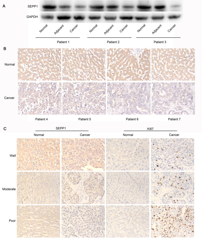
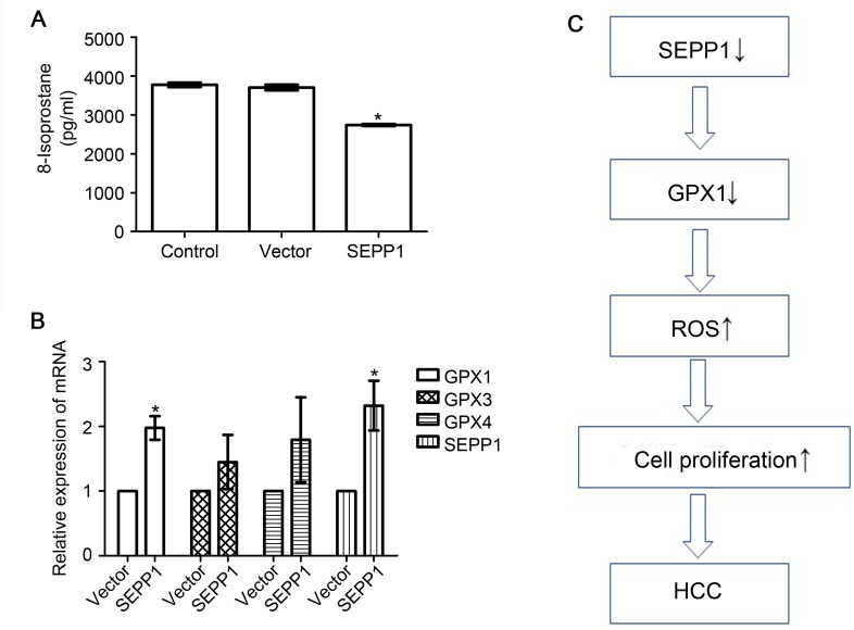
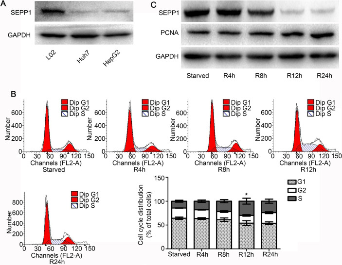
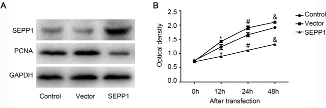
Selenoprotein P (SEPP1) is a kind of secretory glycoproteins with an antioxidant effect during the development of some diseases. In this study, we attempted to observe the expression of SEPP1 in livers from the patients with hepatocellular carcinoma (HCC) and explore its effect on HCC cells. All the tissues from patients with HCC were obtained from Affiliated Hospital of Nantong University. Western blot and immunohistochemical results showed that SEPP1 was reduced in HCC liver tissues. Its expression was negatively correlated with Ki67 expression in tissues. The expression of SEPP1 in normal liver cell line was significantly higher than those in the liver cancer cell lines. Serum starvation and release experiment demonstrated that SEPP1 expression was reduced and PCNA expression was increased, when the serum was re-added into cell culture system and the cells were on a proliferation state. After SEPP1 over-expression plasmid was transfected into HepG2 cells, cell proliferation of HepG2 cells and PCNA expression level were all inhibited by SEPP1. Results obtained via 8-isoprostane ELISA further indicated that inhibited ROS level was found in HepG2 cells transfected with SEPP1 over-expression plasmid. In addition, RT-qPCR results demonstrated that GPX1 expression levels increased in HepG2 cells transfected with SEPP1 over-expression plasmid. In conclusion, SEPP1 may inhibit the proliferation of HCC cells, accompanied by the reduction of ROS production and the increasing of GPX1 expression.
硒蛋白 P(SEPP1)是一种具有抗氧化作用的分泌性糖蛋白,在某些疾病的发展过程中发挥作用。本研究试图观察 SEPP1 在肝癌(HCC)患者肝脏中的表达,并探讨其对 HCC 细胞的影响。所有 HCC 患者的组织均来自南通大学附属医院。Western blot 和免疫组织化学结果表明,SEPP1 在 HCC 肝组织中表达降低。其表达与组织中 Ki67 的表达呈负相关。正常肝细胞系中 SEPP1 的表达明显高于肝癌细胞系。血清饥饿和释放实验表明,当血清重新加入细胞培养系统且细胞处于增殖状态时,SEPP1 的表达减少,PCNA 的表达增加。将 SEPP1 过表达质粒转染至 HepG2 细胞后,SEPP1 抑制了 HepG2 细胞的增殖和 PCNA 表达水平。8-异前列腺素 ELISA 的结果进一步表明,转染 SEPP1 过表达质粒的 HepG2 细胞中 ROS 水平降低。此外,RT-qPCR 结果表明,转染 SEPP1 过表达质粒的 HepG2 细胞中 GPX1 的表达水平升高。综上所述,SEPP1 可能通过降低 ROS 生成和增加 GPX1 的表达来抑制 HCC 细胞的增殖。