Ba Alexandre, Shams Marwa, Schmidt Sabine, Eckstein Miguel P, Verdun Francis R, Bochud François O
Lausanne University Hospital and University of Lausanne, Institute of Radiation Physics, Lausanne, Switzerland.
University of Lausanne, Lausanne, Switzerland.
J Med Imaging (Bellingham). 2020 Jul;7(4):045501. doi: 10.1117/1.JMI.7.4.045501. Epub 2020 Jul 24.
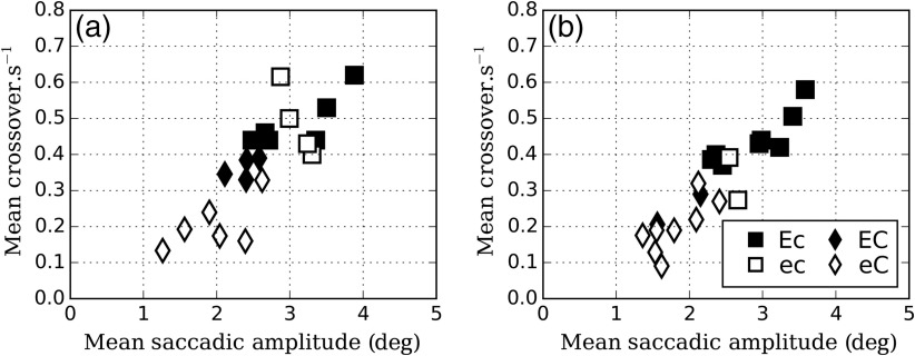
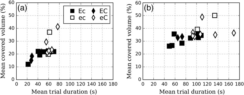
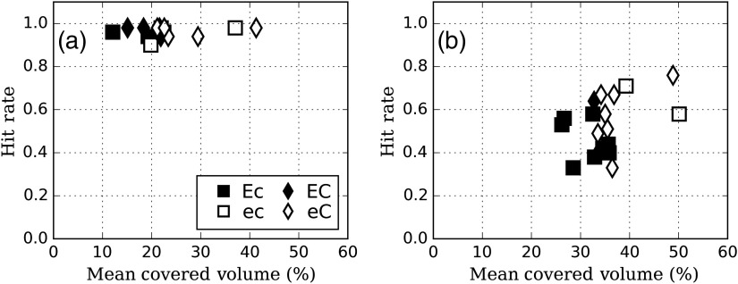
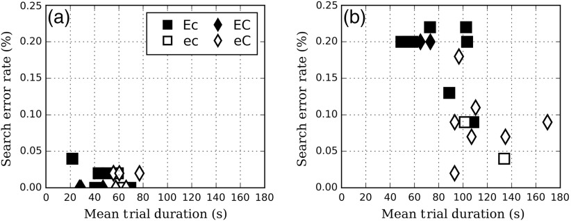
Visual search using volumetric images is becoming the standard in medical imaging. However, we do not fully understand how eye movement strategies mediate diagnostic performance. A recent study on computed tomography (CT) images showed that the search strategies of radiologists could be classified based on saccade amplitudes and cross-quadrant eye movements [eye movement index (EMI)] into two categories: drillers and scanners. We investigate how the number of times a radiologist scrolls in a given direction during analysis of the images (number of courses) could add a supplementary variable to use to characterize search strategies. We used a set of 15 normal liver CT images in which we inserted 1 to 5 hypodense metastases of two different signal contrast amplitudes. Twenty radiologists were asked to search for the metastases while their eye-gaze was recorded by an eye-tracker device (EyeLink1000, SR Research Ltd., Mississauga, Ontario, Canada). We found that categorizing radiologists based on the number of courses (rather than EMI) could better predict differences in decision times, percentage of image covered, and search error rates. Radiologists with a larger number of courses covered more volume in more time, found more metastases, and made fewer search errors than those with a lower number of courses. Our results suggest that the traditional definition of drillers and scanners could be expanded to include scrolling behavior. Drillers could be defined as scrolling back and forth through the image stack, each time exploring a different area on each image (low EMI and high number of courses). Scanners could be defined as scrolling progressively through the stack of images and focusing on different areas within each image slice (high EMI and low number of courses). Together, our results further enhance the understanding of how radiologists investigate three-dimensional volumes and may improve how to teach effective reading strategies to radiology residents.
使用容积图像进行视觉搜索正成为医学成像的标准做法。然而,我们尚未完全了解眼动策略是如何调节诊断性能的。最近一项针对计算机断层扫描(CT)图像的研究表明,放射科医生的搜索策略可根据扫视幅度和跨象限眼动[眼动指数(EMI)]分为两类:钻孔者和扫描者。我们研究了放射科医生在图像分析过程中沿给定方向滚动的次数(行程数)如何能添加一个补充变量来用于表征搜索策略。我们使用了一组15张正常肝脏CT图像,在其中插入了1至5个具有两种不同信号对比幅度的低密度转移灶。20名放射科医生被要求搜索这些转移灶,同时他们的眼睛注视情况由眼动追踪设备(EyeLink1000,SR Research Ltd.,加拿大安大略省密西沙加)记录。我们发现,根据行程数(而非EMI)对放射科医生进行分类能够更好地预测决策时间、图像覆盖百分比和搜索错误率方面的差异。行程数较多的放射科医生比行程数较少的医生在更多时间内覆盖了更大的区域,发现了更多转移灶,并且搜索错误更少。我们的结果表明,钻孔者和扫描者的传统定义可以扩展到包括滚动行为。钻孔者可定义为在图像堆栈中来回滚动,每次在每张图像上探索不同区域(低EMI和高行程数)。扫描者可定义为逐步滚动浏览图像堆栈并专注于每个图像切片内的不同区域(高EMI和低行程数)。总之,我们的结果进一步加深了对放射科医生如何研究三维容积的理解,并可能改进向放射科住院医师教授有效阅读策略的方式。