Hotchkiss Brain Institute, Cumming School of Medicine, University of Calgary, Calgary, AB, Canada.
Department of Radiology, Cumming School of Medicine, University of Calgary, Calgary, AB, Canada.
Sci Rep. 2020 Aug 3;10(1):13007. doi: 10.1038/s41598-020-69991-z.
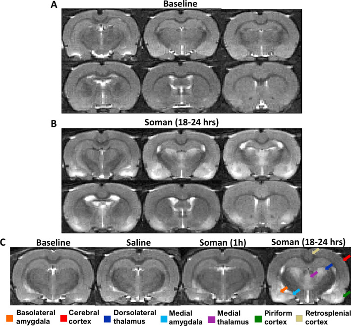
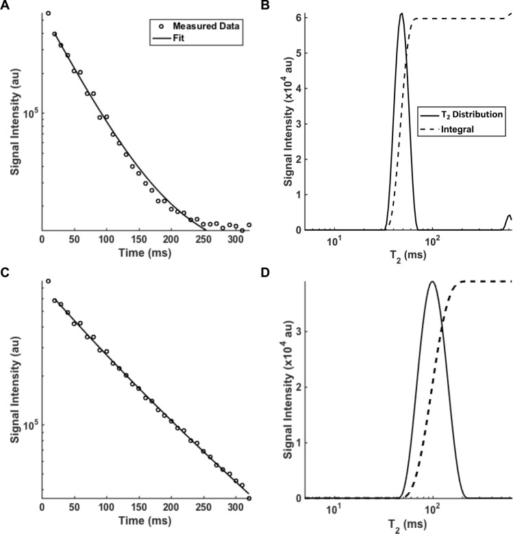
Organophosphorus compounds, such as chemical warfare nerve agents and pesticides, are known to cause neurological damage. This study measured nerve agent-related neuropathology and determined whether quantitative T MRI could be used as a biomarker of neurodegeneration. Quantitative T MRI was performed using a 9.4 T MRI on rats prior to and following soman exposure. T images were taken at least 24 h prior, 1 h and 18-24 h after soman exposure. Rats were pre- and post-treated with HI-6 dimethanesulfonate and atropine methyl nitrate. A multicomponent T acquisition and analysis was performed. Brains were stained with Fluoro-Jade C to assess neurodegeneration. Rats exposed to soman developed behavioral expression of electrographic seizures. At 18-24 h after soman exposure, significant increases in T, a possible marker of edema, were found in multiple regions. The largest changes were in the piriform cortex (before: 47.7 ± 1.4 ms; 18-24 h: 82.3 ± 13.4 ms). Fluoro-Jade C staining showed significant neurodegeneration 18-24 h post exposure. The piriform cortex had the strongest correlation between the change in relaxation rate and percent neurodegeneration (r = 0.96, p < 0.001). These findings indicate there is regionally specific neurodegeneration 24 h after exposure to soman. The high correlation between T relaxivity and histopathology supports the use of T as a marker of injury.
有机磷化合物,如化学战剂神经毒剂和杀虫剂,已知会造成神经损伤。本研究测量了与神经毒剂相关的神经病理学,并确定定量 T MRI 是否可作为神经退行性变的生物标志物。在 soman 暴露前后,使用 9.4 T MRI 对大鼠进行定量 T MRI 检查。在 soman 暴露前至少 24 小时、暴露后 1 小时和 18-24 小时拍摄 T 图像。大鼠用 HI-6 二甲磺酸酯和硝酸甲基阿托品进行预处理和后处理。进行了多分量 T 采集和分析。用 Fluoro-Jade C 染色评估神经退行性变。接触 soman 的大鼠出现电发作的行为表现。在 soman 暴露后 18-24 小时,发现多个区域的 T 值显著增加,T 值可能是水肿的标志物。最大的变化发生在梨状皮层(暴露前:47.7±1.4 ms;18-24 小时:82.3±13.4 ms)。Fluoro-Jade C 染色显示暴露后 18-24 小时有明显的神经退行性变。梨状皮层的弛豫率变化与神经退行性变百分比之间具有最强的相关性(r=0.96,p<0.001)。这些发现表明,暴露于 soman 后 24 小时会出现区域性特异性神经退行性变。T 弛豫率与组织病理学之间的高度相关性支持将 T 用作损伤标志物。