Department of Breast Medical Oncology, The University of Texas MD Anderson Cancer Center, Houston, TX, USA.
Department of Neuroradiology, The University of Texas MD Anderson Cancer Center, Houston, TX, USA.
Br J Cancer. 2020 Oct;123(9):1417-1423. doi: 10.1038/s41416-020-1008-2. Epub 2020 Aug 4.
CNS miliary metastasis (MiM) is poorly recognised in breast and other malignancies. Given its rarity, little epidemiologic, radiographic and clinical data are known. Although usually identified on neuroimaging, criteria for radiographic diagnosis do not exist. In this analysis, we establish its presence in breast cancer and identify factors contributing to outcome.
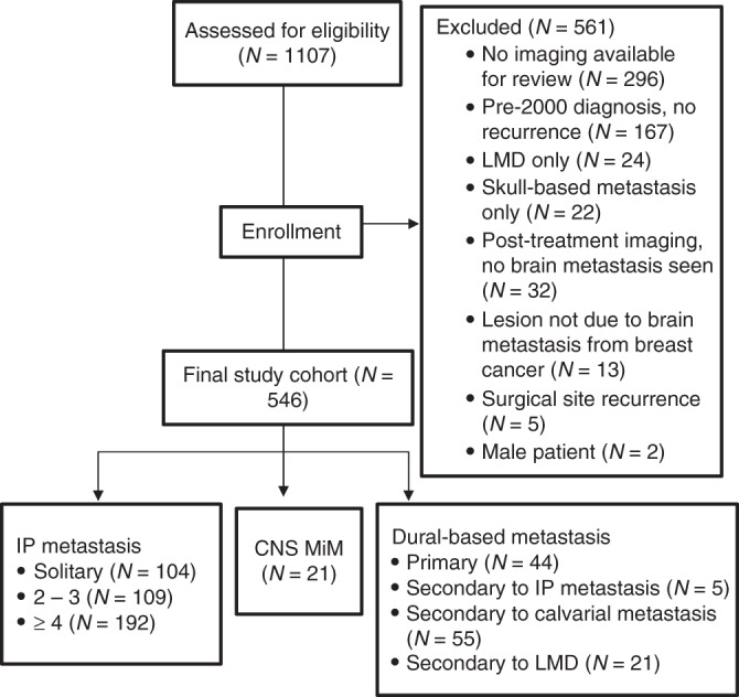
We identified 546 female patients with brain metastasis from breast cancer between 2000 and 2015. Radiographic criteria were established through review of neuroimages by a senior Neuroradiologist, and defined as: (1) ≥20 lesions per image on ≥2 non-contiguous MRI images or ≥10 lesions per image on ≥2 non-contiguous CT images, and (2) bilateral lesions located in both the supratentorial and infratentorial compartments.
Twenty-one MiM cases were identified (3.8%). Number and anatomical distribution of metastases best identified MiM, while lesion size did not. Ten patients were diagnosed with MiM as initial CNS metastasis; 11 developed MiM following known CNS metastasis. Breast cancer subtype did not influence MiM development before or after other CNS metastasis.
This is the first study to propose radiographic criteria for MiM diagnosis. Additional analysis is needed to verify data, but our results may enable a standardised approach for future MiM research.
中枢神经系统(CNS)多发性转移(MiM)在乳腺癌和其他恶性肿瘤中认识不足。由于其罕见性,对其流行病学、影像学和临床数据知之甚少。尽管通常在神经影像学上识别,但不存在放射学诊断标准。在本分析中,我们确定了乳腺癌中 MiM 的存在,并确定了影响预后的因素。
我们在 2000 年至 2015 年间确定了 546 名患有脑转移的女性乳腺癌患者。通过一位资深神经放射科医生对神经图像进行审查,建立了放射学标准,并将其定义为:(1)≥20 个病灶/≥2 个非连续 MRI 图像或≥10 个病灶/≥2 个非连续 CT 图像,以及(2)双侧病灶位于幕上和幕下腔。
确定了 21 例 MiM 病例(3.8%)。转移的数量和解剖分布最能识别 MiM,而病灶大小则不能。10 例患者被诊断为 MiM 作为初始 CNS 转移;11 例患者在已知 CNS 转移后发生 MiM。乳腺癌亚型在前或在后 CNS 转移后并不影响 MiM 的发展。
这是第一项提出 MiM 诊断放射学标准的研究。需要进一步分析来验证数据,但我们的结果可能为未来 MiM 研究提供标准化方法。