Zhang Jinqiang, Zhang Lijuan, Yi Saini, Jiang Xue, Qiao Yan, Zhang Yue, Xiao Chenghong, Zhou Tao
Resource Institute for Chinese & Ethnic Materia Medica, Guizhou University of Traditional Chinese Medicine, Guiyang, China.
School of Life Science and Technology, Center for Informational Biology, University of Electronic Science and Technology of China, Chengdu, China.
Front Cell Neurosci. 2020 Jul 10;14:195. doi: 10.3389/fncel.2020.00195. eCollection 2020.
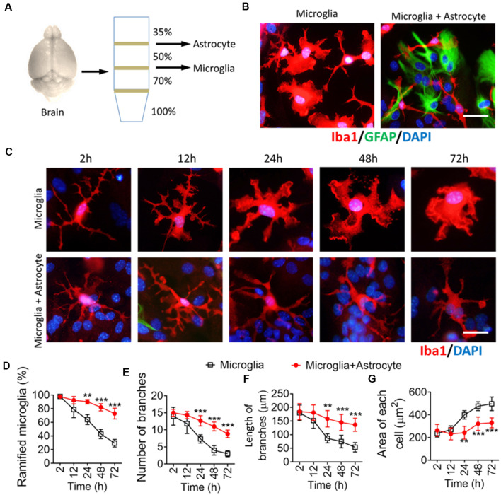
The morphology of microglial cells is often closely related to their functions. The mechanisms that regulate microglial ramification are not well understood. Here we reveal the biological mechanisms by which astrocytes regulate microglial ramification. Morphological variation in mouse microglial cultures was measured in terms of cell area as well as branch number and length. Effects on microglial ramification were analyzed after microinjecting the toxin L-alpha-aminoadipic acid (L-AAA) in the mouse cortex or hippocampus to ablate astrocytes, and after culturing microglia on their own in an astrocyte-conditioned medium (ACM) or together with astrocytes in coculture. TGF-β expression was determined by Western blotting, immunohistochemistry, and ELISA. The TGF-β signaling pathway was blocked by the TGF-β antibody to assess the role of TGF-β on microglial ramification. The results showed that microglia had more and longer branches and smaller cell bodies in brain areas where astrocytes were abundant. In the mouse cortex and hippocampus, ablation of astrocytes by L-AAA decreased number and length of microglial branches and increased the size of cell bodies. Similar results were obtained with isolated microglia in culture. However, isolated microglia were able to maintain their multibranched structure for a long time when cultured on astrocyte monolayers. Ameboid microglia isolated from P0 to P3 mice showed increased ramification when cultured in ACM or on astrocyte monolayers. Microglia cultured on astrocyte monolayers showed more complex branching structures than those cultured in ACM. Blocking astrocyte-derived TGF-β decreased microglial ramification. Astrocytes induced the formation of protuberances on branches of microglia by forming glial fibers that increased traction. These experiments in mice suggest that astrocytes promote microglial ramification by forming glial fibers to create traction and by secreting soluble factors into the surroundings. For example, astrocyte-secreted TGF-β promotes microglia to generate primitive branches, whose ramification is refined by glial fibers.
小胶质细胞的形态通常与其功能密切相关。调节小胶质细胞分支的机制尚未完全明确。在此,我们揭示了星形胶质细胞调节小胶质细胞分支的生物学机制。通过测量细胞面积以及分支数量和长度,来评估小鼠小胶质细胞培养物中的形态变化。在向小鼠皮质或海马中微量注射毒素L-α-氨基己二酸(L-AAA)以清除星形胶质细胞后,以及在星形胶质细胞条件培养基(ACM)中单独培养小胶质细胞或与星形胶质细胞共培养后,分析对小胶质细胞分支的影响。通过蛋白质免疫印迹法、免疫组织化学和酶联免疫吸附测定法测定转化生长因子-β(TGF-β)的表达。使用TGF-β抗体阻断TGF-β信号通路,以评估TGF-β对小胶质细胞分支的作用。结果表明,在星形胶质细胞丰富的脑区,小胶质细胞具有更多、更长的分支以及更小的细胞体。在小鼠皮质和海马中,L-AAA清除星形胶质细胞会减少小胶质细胞分支的数量和长度,并增加细胞体的大小。在培养的分离小胶质细胞中也获得了类似结果。然而,当在星形胶质细胞单层上培养时,分离的小胶质细胞能够长时间维持其多分支结构。从出生后0至3天的小鼠分离出的阿米巴样小胶质细胞,在ACM中或在星形胶质细胞单层上培养时,分支增加。在星形胶质细胞单层上培养的小胶质细胞比在ACM中培养的小胶质细胞表现出更复杂的分支结构。阻断星形胶质细胞衍生的TGF-β会减少小胶质细胞的分支。星形胶质细胞通过形成增加牵引力的神经胶质纤维,诱导小胶质细胞分支上形成突起。这些在小鼠身上进行的实验表明,星形胶质细胞通过形成神经胶质纤维以产生牵引力并向周围环境分泌可溶性因子来促进小胶质细胞的分支。例如,星形胶质细胞分泌的TGF-β促进小胶质细胞产生原始分支,其分支由神经胶质纤维细化。