Christiansen Eric, Singh Nisha, Trahan Amy, Tokman Sofya, Row David, Kalinkin Olga
Department of Radiology, St. Joseph's Hospital and Medical Center, Phoenix, AZ, USA.
Department of Medicine, St. Joseph's Hospital and Medical Center, Phoenix, AZ, USA.
Case Rep Transplant. 2020 Jul 13;2020:5023948. doi: 10.1155/2020/5023948. eCollection 2020.
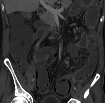
Pneumatosis intestinalis (PI) is a radiologic finding which is characterized by the accumulation of gas within the bowel wall. This radiologic finding is traditionally thought of in the sense of intestinal ischemia. An uncommon cause of this finding is post organ transplantation. We did an institutional and literature review of this finding to demonstrate its distinct imaging features and benign nature. It was observed to occur in approximately 5.2% of patients post lung transplant (23/442). On imaging, it displays an expansile/bubbly appearance of gas within the bowel wall that is distinct from the traditional findings seen in intestinal ischemia. Clinical review showed that posttransplant patients with PI can be successfully managed conservatively with early enteral nutrition, oxygen, antibiotics, and limited follow-up imaging. With the increasing use of organ transplantation, PI is being diagnosed with increased frequency. It is important to let clinicians know of this entity and its potential outcomes.
肠壁积气(PI)是一种放射学表现,其特征为肠壁内气体积聚。传统上,这种放射学表现被认为与肠缺血有关。该表现的一个不常见原因是器官移植后。我们对这一表现进行了机构研究和文献综述,以展示其独特的影像学特征和良性本质。据观察,它在肺移植术后患者中发生率约为5.2%(23/442)。在影像学上,它表现为肠壁内气体呈扩张性/气泡状外观,这与肠缺血的传统表现不同。临床研究表明,移植后发生PI的患者可通过早期肠内营养、吸氧、使用抗生素及有限的随访影像学检查成功进行保守治疗。随着器官移植的日益普及,PI的诊断频率也在增加。让临床医生了解这一实体及其潜在结果很重要。