Théroux-Rancourt Guillaume, Jenkins Matthew R, Brodersen Craig R, McElrone Andrew, Forrestel Elisabeth J, Earles J Mason
Institute of Botany University of Natural Resources and Life Sciences Vienna Austria.
Department of Viticulture and Enology University of California Davis California USA.
Appl Plant Sci. 2020 Jul 31;8(7):e11380. doi: 10.1002/aps3.11380. eCollection 2020 Jul.
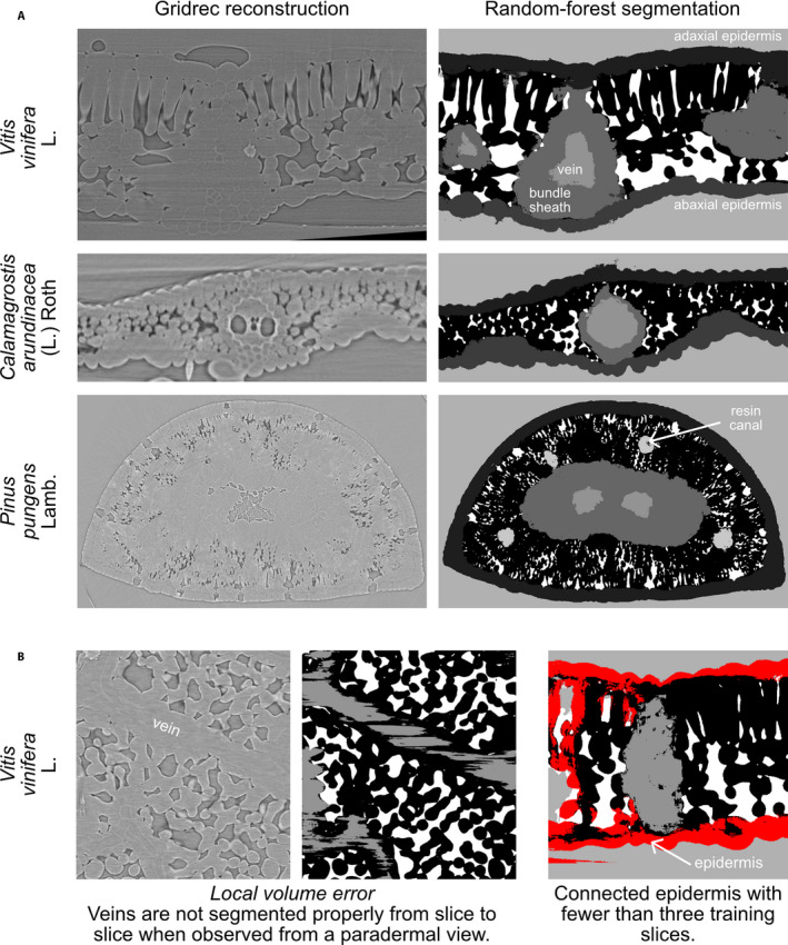
X-ray microcomputed tomography (microCT) can be used to measure 3D leaf internal anatomy, providing a holistic view of tissue organization. Previously, the substantial time needed for segmenting multiple tissues limited this technique to small data sets, restricting its utility for phenotyping experiments and limiting our confidence in the inferences of these studies due to low replication numbers.
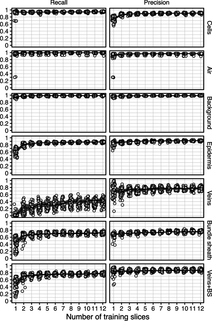
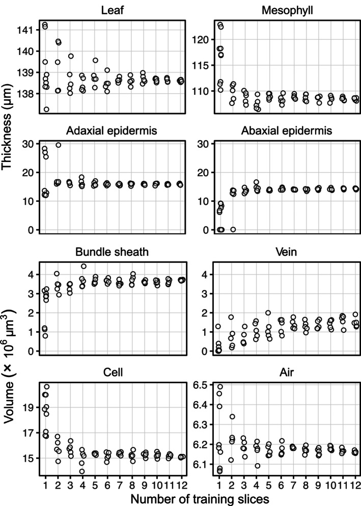
We present a Python codebase for random forest machine learning segmentation and 3D leaf anatomical trait quantification that dramatically reduces the time required to process single-leaf microCT scans into detailed segmentations. By training the model on each scan using six hand-segmented image slices out of >1500 in the full leaf scan, it achieves >90% accuracy in background and tissue segmentation.
Overall, this 3D segmentation and quantification pipeline can reduce one of the major barriers to using microCT imaging in high-throughput plant phenotyping.
X射线显微计算机断层扫描(microCT)可用于测量叶片内部的三维解剖结构,提供组织组织的整体视图。以前,分割多个组织所需的大量时间将该技术限制在小数据集上,限制了其在表型实验中的实用性,并由于复制数量低而限制了我们对这些研究推断的信心。
我们提出了一个用于随机森林机器学习分割和三维叶片解剖特征量化的Python代码库,该代码库显著减少了将单叶microCT扫描处理成详细分割所需的时间。通过在全叶扫描中使用超过1500个手动分割图像切片中的六个对每个扫描进行模型训练,它在背景和组织分割中实现了超过90%的准确率。
总体而言,这种三维分割和量化管道可以减少在高通量植物表型分析中使用microCT成像的一个主要障碍。