Center of Excellence for Regenerative Dentistry and Department of Anatomy, Faculty of Dentistry, Chulalongkorn University, Bangkok, 10330, Thailand.
Oral Biology Research Center, Faculty of Dentistry, Chulalongkorn University, Bangkok, 10330, Thailand.
Sci Rep. 2020 Aug 7;10(1):13329. doi: 10.1038/s41598-020-70277-7.
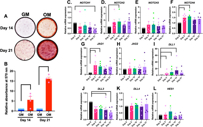
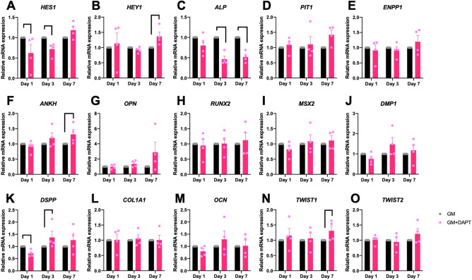
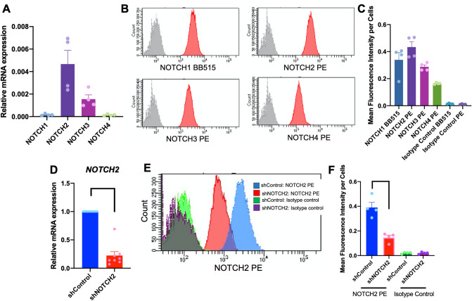
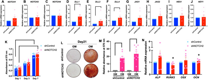
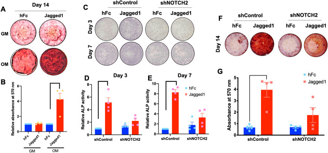
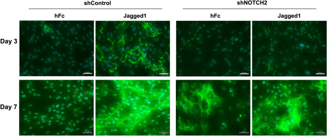
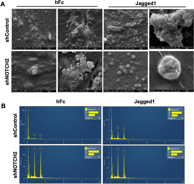
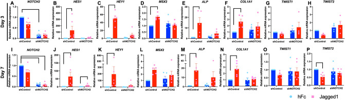
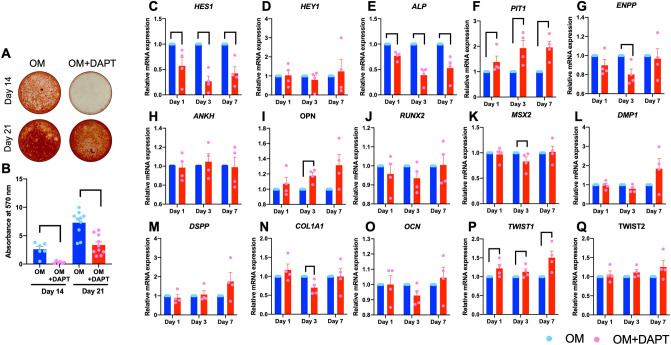
Jagged1 activates Notch signaling and subsequently promotes osteogenic differentiation in human periodontal ligament cells (hPDLs). The present study investigated the participation of the Notch receptor, NOTCH2, in the Jagged1-induced osteogenic differentiation in hPDLs. NOTCH2 and NOTCH4 mRNA expression levels increased during hPDL osteogenic differentiation. However, the endogenous NOTCH2 expression levels were markedly higher compared with NOTCH4. NOTCH2 expression knockdown using shRNA in hPDLs did not dramatically alter their proliferation or osteogenic differentiation compared with the shRNA control. After seeding on Jagged1-immobilized surfaces and maintaining the hPDLs in osteogenic medium, HES1 and HEY1 mRNA levels were markedly reduced in the shNOTCH2-transduced cells compared with the shControl group. Further, shNOTCH2-transduced cells exhibited less alkaline phosphatase enzymatic activity and in vitro mineralization than the shControl cells when exposed to Jagged1. MSX2 and COL1A1 mRNA expression after Jagged1 activation were reduced in shNOTCH2-transduced cells. Endogenous Notch signaling inhibition using a γ-secretase inhibitor (DAPT) attenuated mineralization in hPDLs. DAPT treatment significantly promoted TWIST1, but decreased ALP, mRNA expression, compared with the control. In conclusion, Notch signaling is involved in hPDL osteogenic differentiation. Moreover, NOTCH2 participates in the mechanism by which Jagged1 induced osteogenic differentiation in hPDLs.
Jagged1 激活 Notch 信号通路,随后促进人牙周膜细胞(hPDLs)的成骨分化。本研究探讨了 Notch 受体 NOTCH2 在 Jagged1 诱导的 hPDLs 成骨分化中的参与作用。NOTCH2 和 NOTCH4 mRNA 表达水平在 hPDL 成骨分化过程中增加。然而,内源性 NOTCH2 表达水平明显高于 NOTCH4。用 shRNA 在 hPDLs 中敲低 NOTCH2 表达与 shRNA 对照相比,并没有显著改变其增殖或成骨分化。在 Jagged1 固定表面上接种并将 hPDLs 维持在成骨培养基中后,与 shControl 组相比,shNOTCH2 转导细胞中的 HES1 和 HEY1 mRNA 水平明显降低。此外,与 shControl 细胞相比,暴露于 Jagged1 时,shNOTCH2 转导细胞的碱性磷酸酶酶活性和体外矿化能力较低。Jagged1 激活后 MSX2 和 COL1A1 mRNA 表达在 shNOTCH2 转导细胞中减少。用 γ-分泌酶抑制剂(DAPT)抑制内源性 Notch 信号通路减弱了 hPDLs 的矿化。与对照组相比,DAPT 处理显著促进了 TWIST1,但降低了 ALP 的 mRNA 表达。总之,Notch 信号通路参与 hPDL 成骨分化。此外,NOTCH2 参与了 Jagged1 诱导 hPDLs 成骨分化的机制。