Bobetsis Yiorgos A, Kotsikoris Ioannis, Liapis Christos D, Liasis Nikolaos, Kakisis John, Kourlaba Georgia, Lazari Paraskevi, Antonopoulos Constantine N, Deliargyris Efthymios N, Madianos Phoebus N
Dept of Periodontology, School of Dentistry, National and Kapodistrian University of Athens, Greece.
Dept of Vascular Surgery, Red Cross Hospital, Athens, Greece.
Int J Cardiol Heart Vasc. 2020 Aug 9;30:100601. doi: 10.1016/j.ijcha.2020.100601. eCollection 2020 Oct.
Periodontal disease (PD) is a chronic inflammatory oral condition with potentially important systemic sequelae. We sought to determine whether the presence of PD in patients with severe carotid disease was associated with morphological features consistent with carotid plaque instability.
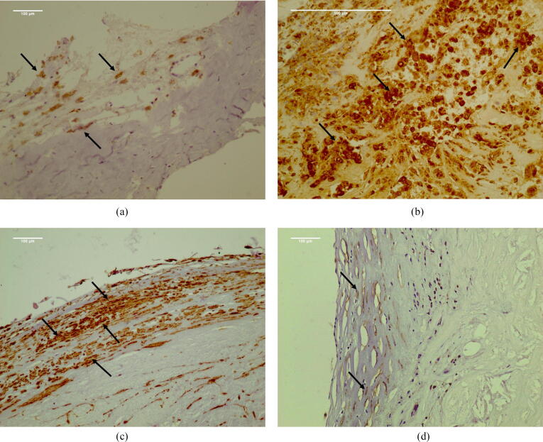
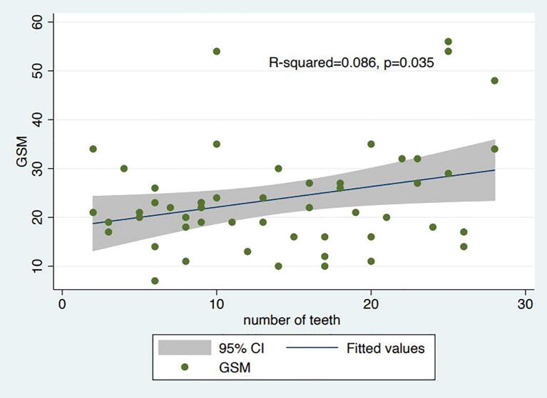
A total of 52 dentate patients hospitalized for carotid endarterectomy (CEA) had standardized assessments of their periodontal status, including measurements of probing pocket depth (PPD), clinical attachment level (CAL) and bleeding on probing (BoP). Carotid plaque morphology was assessed by ultrasound using the gray scale median (GSM) score and by immunohistochemistry using anti-CD68 and anti-alpha-actin antibodies, markers for macrophages and smooth muscle cells (SMCs) respectively.
In total 30/52 patients (58%) had PD. Significant associations were noted between low GSM on ultrasound and each mm in PPD (p = 0.001), each mm in CAL (p = 0.002) and with a 10% increase in BoP (p = 0.009). Using the standardized PERIO definition the association remained robust (aOR = 10.4 [95% CI:2.3-46.3], p = .002). Significant associations were also observed with high macrophage accumulation and each individual PD measure (p < 0.01 for PPD, CAL and BoP) and with the PERIO definition (aOR = 15 [95% CI:1.8-127.8], p = .01). Similarly, low SMC density was also significantly associated with individual measures of PD (p < 0.05 for PPD, CAL and BoP), but not with the PERIO definition (aOR 3.4 [95% CI:0.9-12.8], p = .07).
The presence of PD was significantly associated with both ultrasound and immunohistochemistry features of carotid plaque instability in patients undergoing CEA.
牙周病(PD)是一种慢性炎症性口腔疾病,可能具有重要的全身后遗症。我们试图确定重度颈动脉疾病患者中牙周病的存在是否与颈动脉斑块不稳定的形态学特征相关。
共有52例因颈动脉内膜切除术(CEA)住院的有牙患者接受了牙周状况的标准化评估,包括探诊深度(PPD)、临床附着水平(CAL)和探诊出血(BoP)测量。通过超声使用灰度中位数(GSM)评分评估颈动脉斑块形态,并通过免疫组织化学使用抗CD68和抗α-肌动蛋白抗体分别评估巨噬细胞和平滑肌细胞(SMC)的标志物。
总共30/52例患者(58%)患有牙周病。超声检查中低GSM与PPD每增加1mm(p = 0.001)、CAL每增加1mm(p = 0.002)以及BoP增加10%(p = 0.009)之间存在显著关联。使用标准化的牙周病定义,这种关联仍然很强(调整后比值比[aOR]=10.4[95%置信区间:2.3 - 46.3],p = 0.002)。在巨噬细胞高度聚集与每项牙周病测量指标之间也观察到显著关联(PPD、CAL和BoP的p < 0.01),以及与牙周病定义之间的关联(aOR = 15[95%置信区间:1.8 - 127.8],p = 0.01)。同样,低SMC密度也与牙周病的各项测量指标显著相关(PPD、CAL和BoP的p < 0.05),但与牙周病定义无关(aOR 3.4[95%置信区间:0.9 - 12.8],p = 0.07)。
在接受CEA的患者中,牙周病的存在与颈动脉斑块不稳定的超声和免疫组织化学特征均显著相关。