Department of Ophthalmology, Bundang Jesaeng General Hospital, Daejin Medical Center, Seongnam, Korea.
Department of Neuropsychiatry, Seoul National University College of Medicine, Seoul National University Bundang Hospital, Seongnam, Korea.
Transl Vis Sci Technol. 2020 Jun 17;9(7):17. doi: 10.1167/tvst.9.7.17. eCollection 2020 Jun.
The purpose of this study was to investigate the relationship between the lamina cribrosa (LC) thickness (LCT) as assessed using enhanced depth-imaging (EDI) optical coherence tomography (OCT) and cognitive function in primary open-angle glaucoma (POAG).
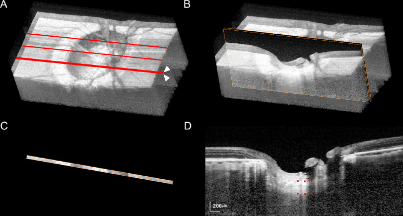
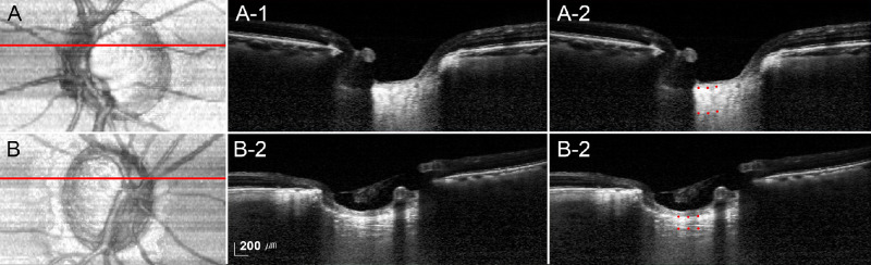
The study consisted of 105 POAG eyes and 23 nonglaucomatous control eyes that completed neuropsychological tests. The optic nerve heads of the patients were imaged using EDI-OCT. B-scan images were constructed in three dimensions using maximum intensity projection (MIP), and the LCT was measured using the thin-slab MIP images. A comprehensive battery consisting of 15 neuropsychological tests was used to evaluate cognitive function.
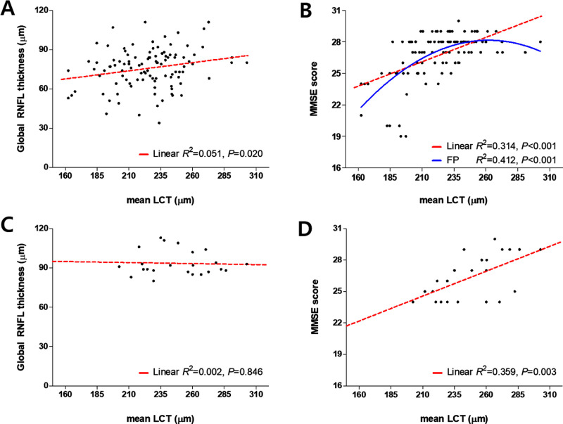
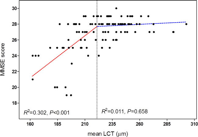
POAG eyes had smaller mean LCT as compared with control eyes ( < 0.001). Age and Mini Mental State Examination (MMSE) scores did not differ between the two groups. Linear regression analysis revealed that lower scores on the MMSE ( < 0.001), presence of glaucoma ( = 0.006), and a smaller global retinal nerve fiber layer thickness ( < 0.001) were independently associated with a smaller mean LCT. Davies' test revealed a statistically significant breakpoint for the mean LCT (221.14 µm), below which a smaller MMSE score was significantly associated with a smaller mean LCT. In POAG eyes with a mean LCT smaller than the breakpoint (< 221.14 µm), not only the global cognition but also the visuospatial function and visual memory were worse than in those with a larger mean LCT (all ≤ 0.003).
Impairment of cognitive function was observed in patients with POAG with a thinner LC. The role of LC imaging as a potential biomarker to monitor cognitive impairment needs further investigation.
LC thinning may reflect a shared mechanism of neurodegenerative diseases in the brain and optic nerve.
本研究旨在探讨使用增强深度成像(EDI)光学相干断层扫描(OCT)评估的视盘筛板(LC)厚度(LCT)与原发性开角型青光眼(POAG)认知功能之间的关系。
本研究纳入了 105 只 POAG 眼和 23 只非青光眼对照眼,所有患者均完成了神经心理学测试。使用 EDI-OCT 对患者的视神经头部进行成像。使用最大强度投影(MIP)构建 B 扫描的三维图像,并使用薄切片 MIP 图像测量 LCT。使用包含 15 个神经心理学测试的综合测试套件评估认知功能。
POAG 眼的平均 LCT 明显小于对照组( < 0.001)。两组的年龄和简易精神状态检查(MMSE)评分无差异。线性回归分析显示,MMSE 评分较低( < 0.001)、存在青光眼( = 0.006)和全视网膜神经纤维层厚度较小( < 0.001)与平均 LCT 较小独立相关。Davies 检验显示平均 LCT 存在统计学上的显著断点(221.14 µm),低于该值时,MMSE 评分较低与平均 LCT 较小显著相关。在 LCT 平均值小于断点(< 221.14 µm)的 POAG 眼中,不仅整体认知功能,而且视空间功能和视觉记忆均较 LCT 较大者差(均 ≤ 0.003)。
在 LC 较薄的 POAG 患者中观察到认知功能受损。LC 成像作为监测认知障碍的潜在生物标志物的作用需要进一步研究。