Wang Zihao, Zhou Xinbo, Xu Yuru, Fan Shiyong, Tian Ning, Zhang Wenyuan, Sheng Fugeng, Lin Jian, Zhong Wu
Beijing Institute of Pharmacology and Toxicology, No. 27 Taiping Road, Beijing 100850, China.
Chinese People's Liberation Army Hospital 307, 8 East Street, Fengtai District, Beijing 100071, China.
ACS Omega. 2020 Aug 7;5(32):20653-20663. doi: 10.1021/acsomega.0c03073. eCollection 2020 Aug 18.
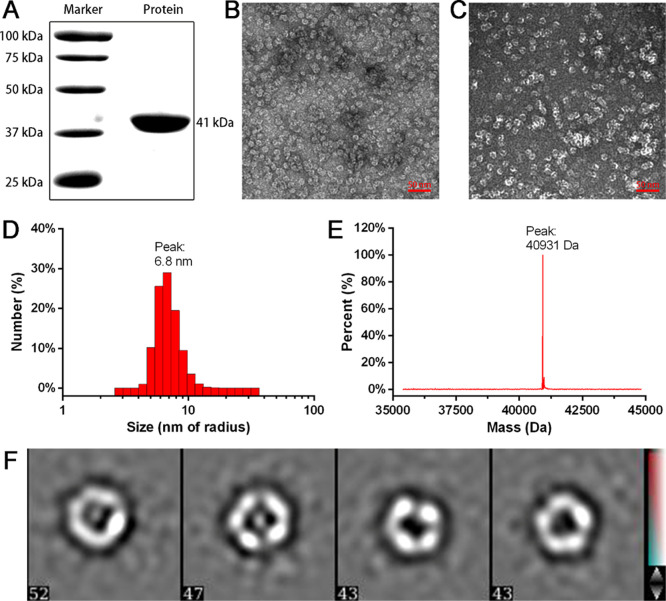
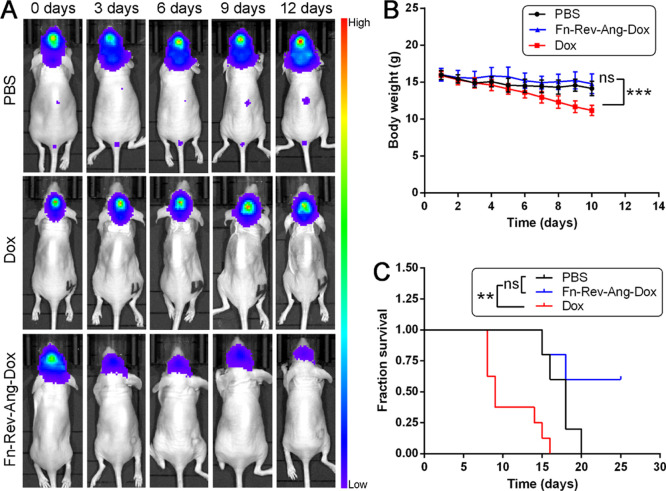
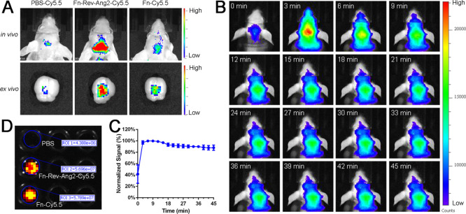
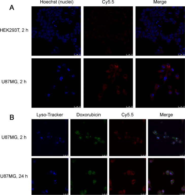
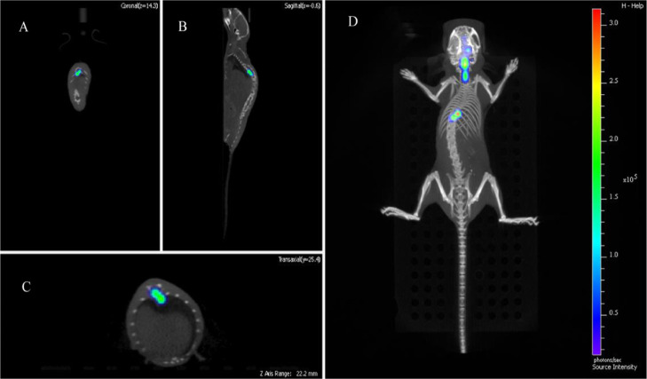
Clinically diagnosing low-grade gliomas and microscopic metastatic tumors in the spinal cord using magnetic resonance imaging (MRI) is challenging, as the blood-brain barrier (BBB) almost completely excludes the MRI contrast agent gadopentetate dimeglumine, GdDTPA (Magnevist), from the brain. The development of a more efficient, safe, and broad-spectrum glioma diagnosis and treatment would therefore have a great clinical value. Based on the high expression levels of both transferrin receptor 1 (TfR1) and low-density lipoprotein receptor-related protein 1 (LRP1) in BBB-related cells and glioma cells, we designed a novel protein nanoparticle, ferritin-HREV107-Angiopep-2 (Fn-Rev-Ang). We found that Fn-Rev-Ang rapidly crossed the BBB in mice and had drug-loading properties. Moreover, the brain MRI signal intensity ratio associated with Fn-Rev-Ang-GdDTPA was higher than that associated with Fn-GdDTPA alone. Importantly, gliomas with diameters below 1 mm and microscopic metastatic tumors in the spinal cord were successfully detected in mice by MRI with Fn-Rev-Ang-GdDTPA, which is not possible using the current clinical MRI technology. In addition, Fn-Rev-Ang-loaded doxorubicin had a strong inhibitory effect on mouse brain gliomas and their metastasis, which significantly prolonged the animal survival time. Thus, our newly constructed Fn-Rev-Ang nanodelivery carrier may help expand the use of MRI to the early diagnosis and treatment of microscopic tumors, thereby offering a possible basis for improving the survival rate of patients with gliomas and microscopic spinal metastatic tumors.
利用磁共振成像(MRI)对脊髓中的低度胶质瘤和微小转移瘤进行临床诊断具有挑战性,因为血脑屏障(BBB)几乎完全阻止MRI造影剂钆喷酸葡胺(GdDTPA,商品名马根维显)进入大脑。因此,开发一种更高效、安全且广谱的胶质瘤诊断和治疗方法将具有巨大的临床价值。基于转铁蛋白受体1(TfR1)和低密度脂蛋白受体相关蛋白1(LRP1)在血脑屏障相关细胞和胶质瘤细胞中的高表达水平,我们设计了一种新型蛋白质纳米颗粒,即铁蛋白-HREV107-血管活性肠肽-2(Fn-Rev-Ang)。我们发现Fn-Rev-Ang能在小鼠体内快速穿过血脑屏障并具有载药特性。此外,与Fn-Rev-Ang-GdDTPA相关的脑MRI信号强度比高于单独使用Fn-GdDTPA时的信号强度比。重要的是,通过使用Fn-Rev-Ang-GdDTPA进行MRI检查,成功在小鼠体内检测到了直径小于1 mm的胶质瘤和脊髓中的微小转移瘤,而目前的临床MRI技术无法做到这一点。此外,负载阿霉素的Fn-Rev-Ang对小鼠脑胶质瘤及其转移具有强烈的抑制作用,显著延长了动物的生存时间。因此,我们新构建的Fn-Rev-Ang纳米递送载体可能有助于将MRI的应用扩展到微小肿瘤的早期诊断和治疗,从而为提高胶质瘤和微小脊髓转移瘤患者的生存率提供可能的依据。