Department of Systems Biology, Columbia University Irving Medical Center, New York, United States.
Medical Scientist Training Program, Columbia University Irving Medical Center, New York, United States.
Elife. 2020 Aug 26;9:e60048. doi: 10.7554/eLife.60048.
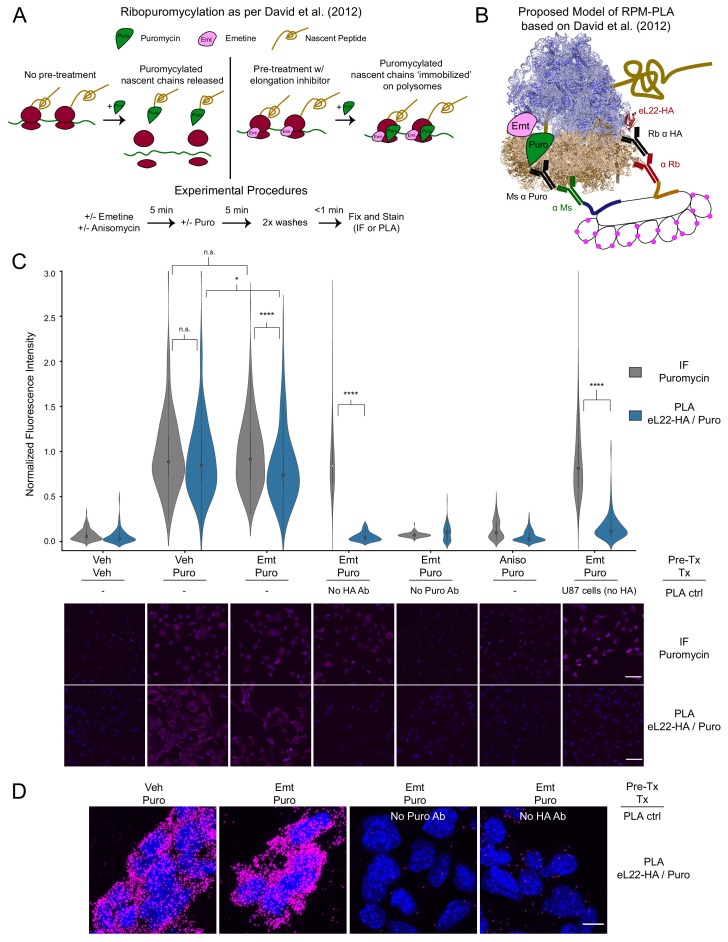
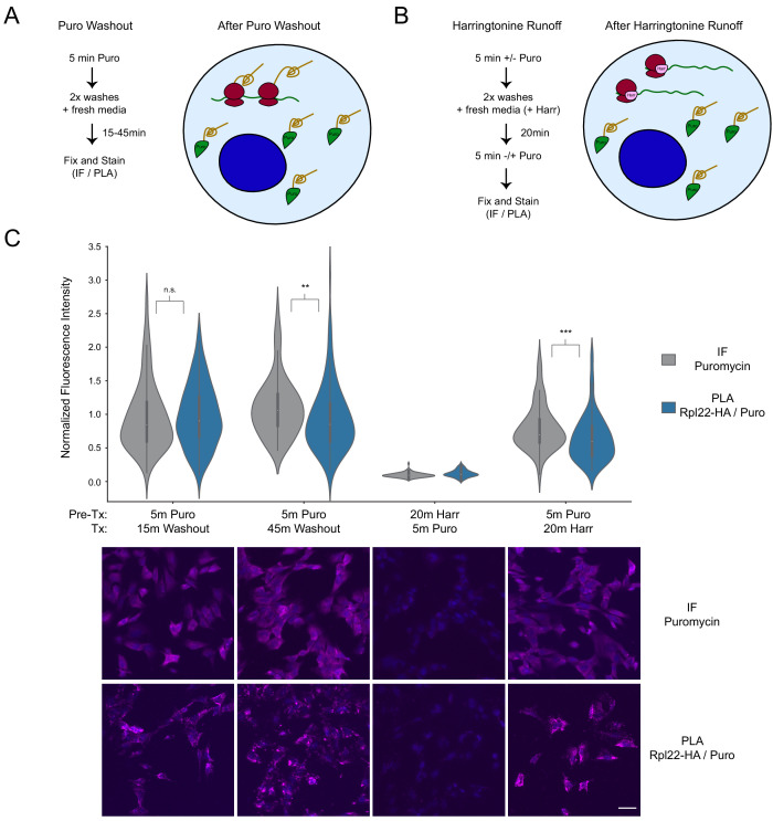
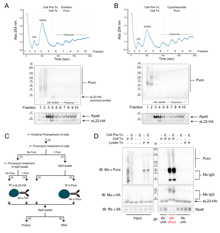
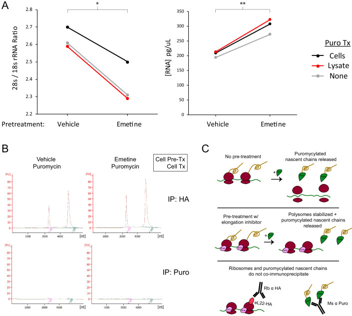
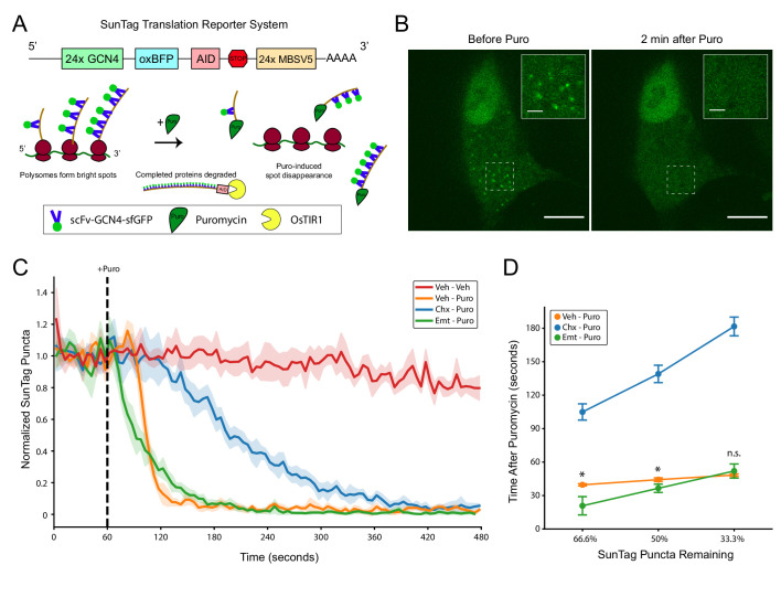
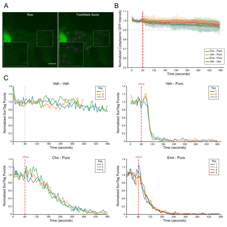
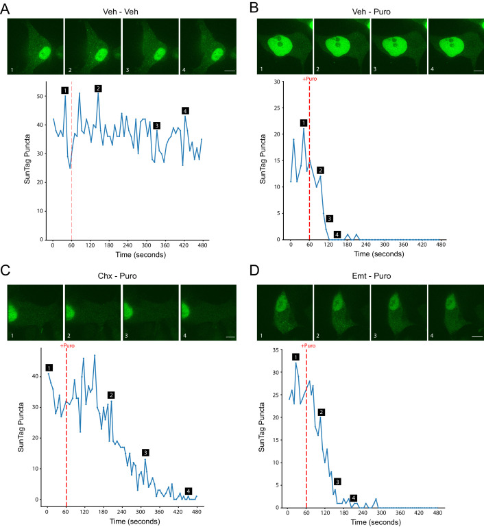
Puromycin is an amino-acyl transfer RNA analog widely employed in studies of protein synthesis. Since puromycin is covalently incorporated into nascent polypeptide chains, anti-puromycin immunofluorescence enables visualization of nascent protein synthesis. A common assumption in studies of local messenger RNA translation is that the anti-puromycin staining of puromycylated nascent polypeptides in fixed cells accurately reports on their original site of translation, particularly when ribosomes are stalled with elongation inhibitors prior to puromycin treatment. However, when we attempted to implement a proximity ligation assay to detect ribosome-puromycin complexes, we found no evidence to support this assumption. We further demonstrated, using biochemical assays and live cell imaging of nascent polypeptides in mammalian cells, that puromycylated nascent polypeptides rapidly dissociate from ribosomes even in the presence of elongation inhibitors. Our results suggest that attempts to define precise subcellular translation sites using anti-puromycin immunostaining may be confounded by release of puromycylated nascent polypeptide chains prior to fixation.
嘌呤霉素是一种广泛应用于蛋白质合成研究的氨酰基 tRNA 类似物。由于嘌呤霉素通过共价键掺入新生多肽链中,因此抗嘌呤霉素免疫荧光可用于观察新生蛋白质的合成。在局部信使 RNA 翻译研究中,一个常见的假设是,用嘌呤霉素处理固定细胞中的嘌呤霉素化新生多肽的抗嘌呤霉素染色准确地反映了它们最初的翻译位置,特别是当核糖体在嘌呤霉素处理前被伸长抑制剂阻止时。然而,当我们试图实施邻近连接测定来检测核糖体-嘌呤霉素复合物时,我们没有发现任何证据支持这一假设。我们进一步通过生化测定和哺乳动物细胞中新生多肽的活细胞成像证明,即使存在伸长抑制剂,嘌呤霉素化的新生多肽也会迅速从核糖体上解离。我们的结果表明,使用抗嘌呤霉素免疫染色来定义精确的亚细胞翻译位点的尝试可能会因固定前释放嘌呤霉素化的新生多肽链而受到干扰。