Sapa-Wojciechowska Agnieszka, Rak-Pasikowska Alina, Pormańczuk Kornel, Czapla Bartłomiej, Bil-Lula Iwona
Division of Clinical Chemistry and Laboratory Hematology, Department of Medical Laboratory Diagnostics, Faculty of Pharmacy, Wroclaw Medical University, 50-556 Wroclaw, Poland.
Department of Surgery, 4th Military Teaching Hospital in Wroclaw, 50-981 Wroclaw, Poland.
Cardiol Res Pract. 2020 Aug 12;2020:9036157. doi: 10.1155/2020/9036157. eCollection 2020.
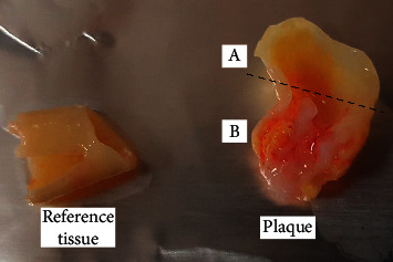
20 patients undergoing routine carotid endarterectomy and 40 healthy volunteers were enrolled in this study. MMPs activity and OPG and FN concentrations were measured in atherosclerotic plaques and nonchanged contiguous tissue after homogenization as well as in plasma from patients and reference group. The activity of MMPs was evaluated by gelatin zymography, and the concentration of OPG and FN was assessed by ELISA.
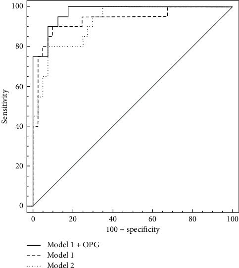
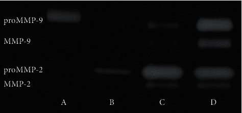
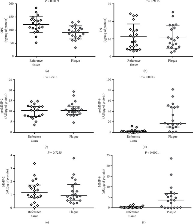
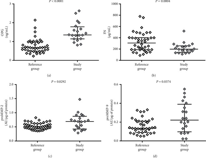
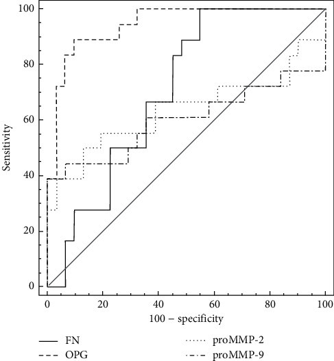
OPG concentration and MMP-9 activity showed differences between plaque and nonchanged tissue; OPG was higher in adjacent tissue (=0.0009), whereas MMP-9 was higher in plaque (proMMP-9 =0.0003; MMP-9 < 0.0001). The OPG plasma concentration and both MMPs plasma activity were higher in patients (OPG < 0.001; proMMP-2 =0.0292; and proMMP-9 =0.0374), while FN plasma concentration was lower in patients than in the reference group (=0.0004). The ROC curves analysis showed the highest AUC for OPG (0.943) with 85.0% sensitivity and 92.1% specificity.
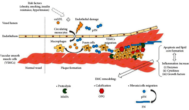
The atherosclerotic plaque and the contiguous artery wall are biochemically different. OPG shows the highest potential to be a marker of advanced carotid atherosclerosis.
本研究纳入了20例行常规颈动脉内膜切除术的患者和40名健康志愿者。在匀浆后,测定动脉粥样硬化斑块及相邻未病变组织中的基质金属蛋白酶(MMPs)活性、骨保护素(OPG)和纤连蛋白(FN)浓度,同时测定患者组和参照组血浆中的上述指标。通过明胶酶谱法评估MMPs的活性,采用酶联免疫吸附测定法评估OPG和FN的浓度。
OPG浓度和MMP-9活性在斑块组织和未病变组织之间存在差异;相邻组织中的OPG较高(=0.0009),而斑块中的MMP-9较高(前MMP-9 =0.0003;MMP-9 <0.0001)。患者血浆中OPG浓度以及两种MMPs的血浆活性均较高(OPG <0.001;前MMP-2 =0.0292;前MMP-9 =0.0374),而患者血浆中FN浓度低于参照组(=0.0004)。ROC曲线分析显示,OPG的曲线下面积(AUC)最高(0.943),敏感性为85.0%,特异性为92.1%。
动脉粥样硬化斑块与相邻动脉壁在生化方面存在差异。OPG最有可能成为晚期颈动脉粥样硬化的标志物。