Institute for Clinical Molecular MRI in the Musculoskeletal System, Karl Landsteiner Society, Vienna, Austria.
High-Field MR Center, Department of Biomedical Imaging and Image-guided Therapy, Medical University of Vienna, Vienna, Austria.
J Magn Reson Imaging. 2021 Jul;54(1):58-75. doi: 10.1002/jmri.27326. Epub 2020 Aug 26.
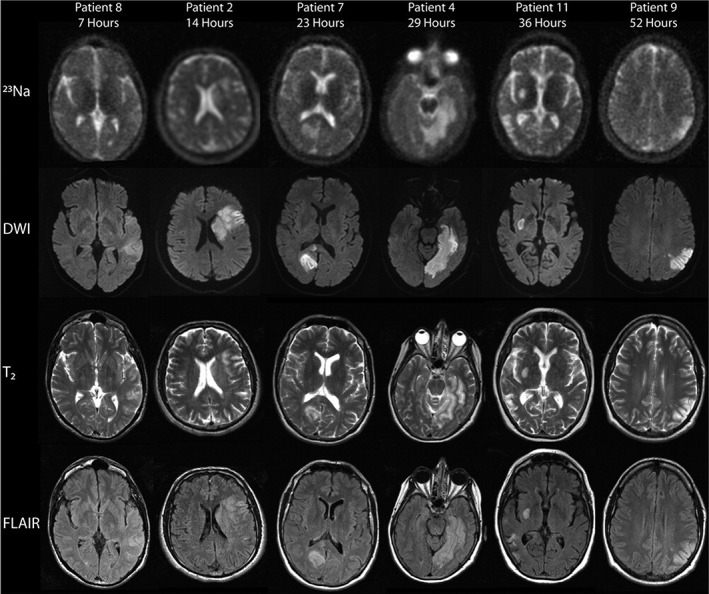
Sodium magnetic resonance imaging ( Na-MRI) is a highly promising imaging modality that offers the possibility to noninvasively quantify sodium content in the tissue, one of the most relevant parameters for biochemical investigations. Despite its great potential, due to the intrinsically low signal-to-noise ratio (SNR) of sodium imaging generated by low in vivo sodium concentrations, low gyromagnetic ratio, and substantially shorter relaxation times than for proton ( H) imaging, Na-MRI is extremely challenging. In this article, we aim to provide a comprehensive overview of the literature that has been published in the last 10-15 years and which has demonstrated different technical designs for a range of Na-MRI methods applicable for disease diagnoses and treatment efficacy evaluations. Currently, a wider use of 3.0T and 7.0T systems provide imaging with the expected increase in SNR and, consequently, an increased image resolution and a reduced scanning time. A great interest in translational research has enlarged the field of sodium MRI applications to almost all parts of the body: articular cartilage tendons, spine, heart, breast, muscle, kidney, and brain, etc., and several pathological conditions, such as tumors, neurological and degenerative diseases, and others. The quantitative parameter, tissue sodium concentration, which reflects changes in intracellular sodium concentration, extracellular sodium concentration, and intra-/extracellular volume fractions is becoming acknowledged as a reliable biomarker. Although the great potential of this technique is evident, there must be steady technical development for Na-MRI to become a standard imaging tool. The future role of sodium imaging is not to be considered as an alternative to H MRI, but to provide early, diagnostically valuable information about altered metabolism or tissue function associated with disease genesis and progression. LEVEL OF EVIDENCE: 1 TECHNICAL EFFICACY STAGE: 1.
钠磁共振成像(Na-MRI)是一种极具前景的成像方式,它提供了一种非侵入性的方法来定量组织中的钠含量,这是生物化学研究中最相关的参数之一。尽管具有巨大的潜力,但由于体内钠浓度低、磁旋比低以及弛豫时间比质子(H)成像短得多,导致钠成像的固有信噪比(SNR)非常低,因此 Na-MRI 极具挑战性。本文旨在对过去 10-15 年发表的文献进行全面综述,这些文献展示了一系列适用于疾病诊断和治疗效果评估的 Na-MRI 方法的不同技术设计。目前,3.0T 和 7.0T 系统的广泛应用为成像提供了预期的 SNR 提高,从而提高了图像分辨率和降低了扫描时间。对转化研究的浓厚兴趣将钠磁共振成像的应用领域扩大到了身体的几乎所有部位:关节软骨、肌腱、脊柱、心脏、乳房、肌肉、肾脏和大脑等,以及许多病理状况,如肿瘤、神经退行性疾病等。定量参数,即组织钠浓度,反映了细胞内钠浓度、细胞外钠浓度和细胞内外体积分数的变化,正在被认为是一种可靠的生物标志物。尽管这项技术具有巨大的潜力,但 Na-MRI 要成为一种标准的成像工具,必须要有稳定的技术发展。钠成像的未来作用不是替代 H MRI,而是提供与疾病发生和发展相关的代谢或组织功能改变的早期、具有诊断价值的信息。证据水平:1 技术功效阶段:1。