The Key Laboratory of Cardiovascular Remodeling and Function Research, Chinese Ministry of Education, Chinese National Health Commission and Chinese Academy of Medical Sciences, The State and Shandong Province Joint Key Laboratory of Translational Cardiovascular Medicine, Department of Cardiology, Qilu Hospital, Cheeloo College of Medicine, Shandong University, Jinan, China.
Department of Cardiology, Shandong Provincial The First Affiliated Hospital of Shandong First Medical University, Jinan, China.
J Cell Mol Med. 2020 Oct;24(20):11729-11741. doi: 10.1111/jcmm.15783. Epub 2020 Aug 27.
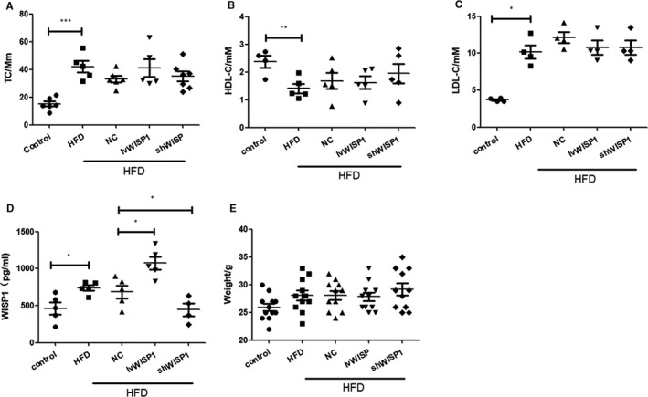
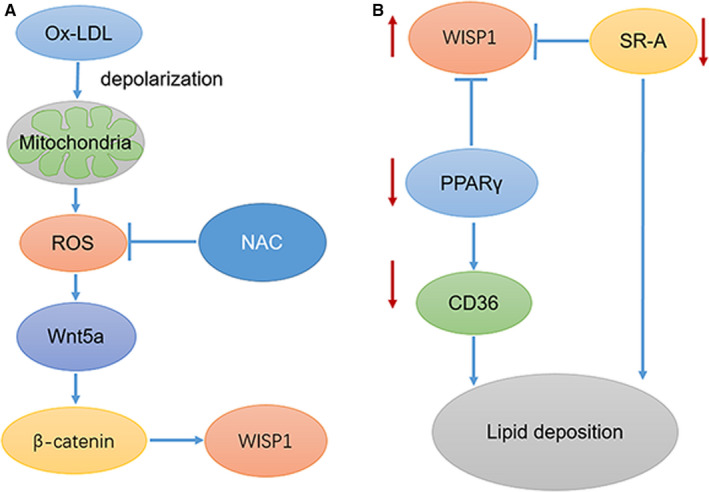
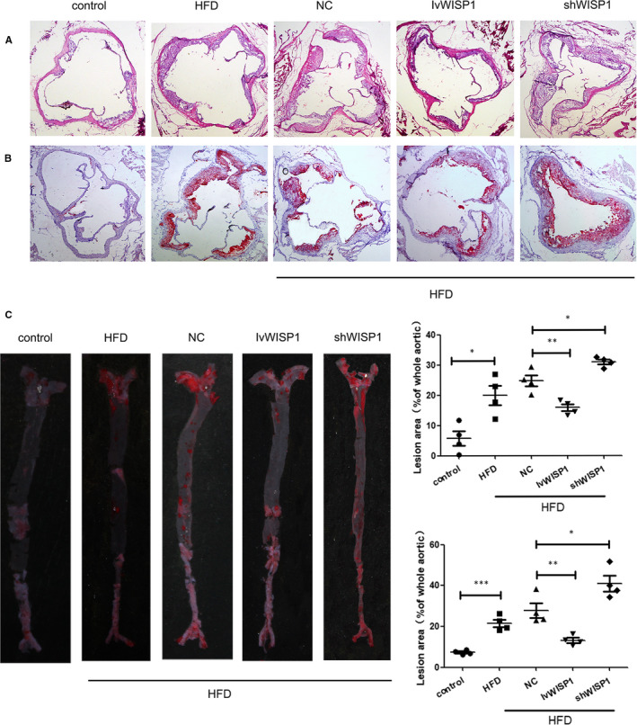
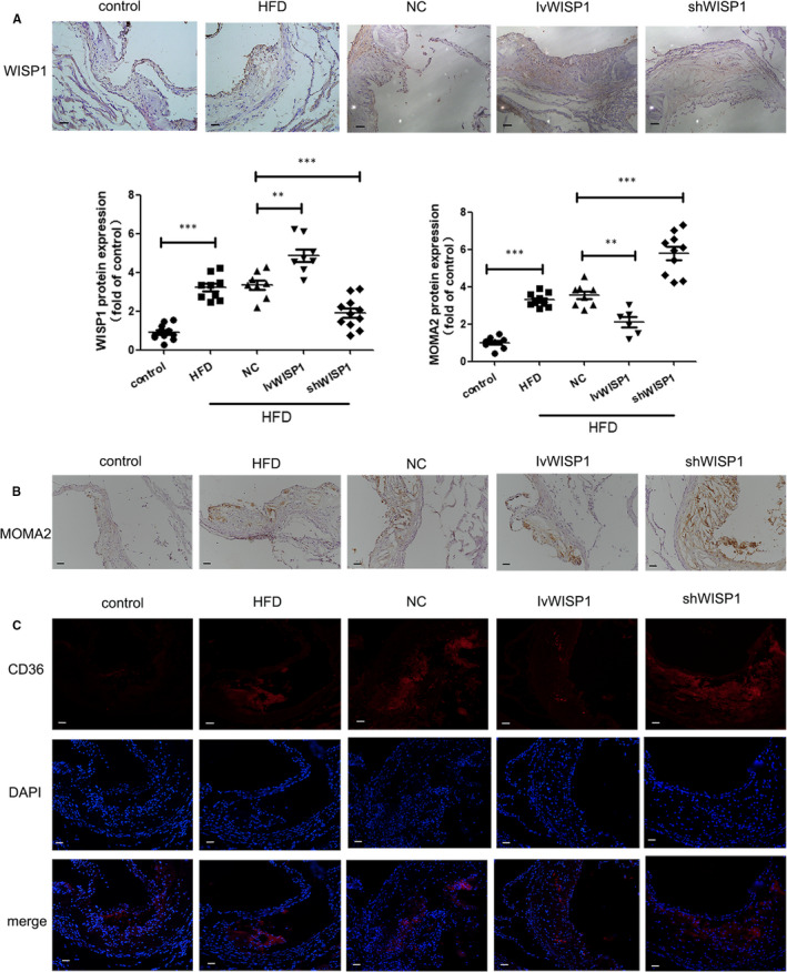
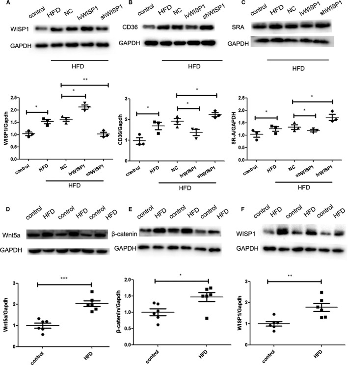
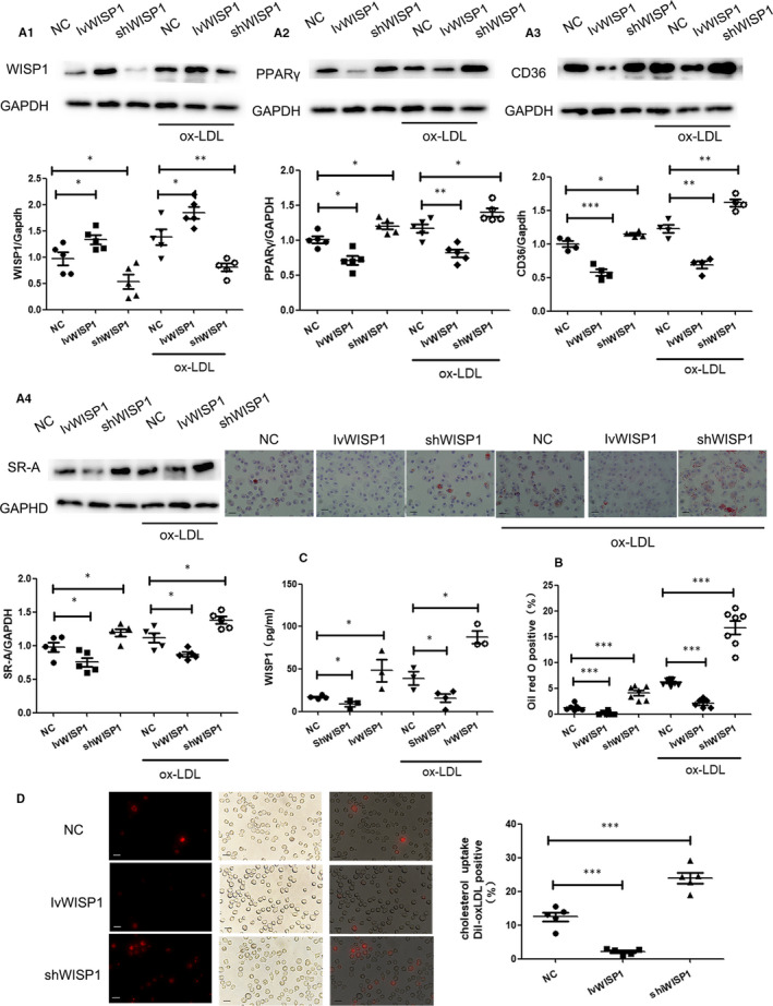
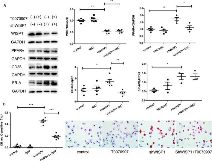
Lipid deposition in macrophages plays an important role in atherosclerosis. The WNT1-inducible signalling pathway protein 1(WISP1) can promote proliferation and migration of smooth muscle cells. Its expression is up-regulated in obesity, which is associated with atherosclerosis, but the effect of WISP1 on atherosclerosis remains unclear. Thus, the objective of our study was to elucidate the role of WISP and its mechanism of action in atherosclerosis via in vivo and in vitro experiments. In our experiment, ApoE-/- mice were divided into 5 groups: control, high-fat diet (HFD), null lentivirus (HFD + NC), lentivirus WISP1 (HFD + IvWISP1) and WISP1-shRNA (HFD + shWISP1). Oil Red O staining, immunofluorescence and immunohistochemistry of the aortic sinuses were conducted. Macrophages (RAW264.7 cell lines and peritoneal macrophages) were stimulated with 50 μg/mL oxidized low-density lipoprotein (ox-LDL); then, the reactive oxygen species (ROS) level was measured. Oil Red O staining and Dil-ox-LDL (ox-LDL with Dil dye) uptake measurements were used to test lipid deposition of peritoneal macrophages. WISP1, CD36, SR-A and PPARγ expression levels were measured via Western blotting and ELISA. The results showed that HFD mice had increased WISP1, CD36 and SR-A levels. The plaque lesion area increased when WISP1 was down-regulated, and lipid uptake and foam cell formation were inhibited when WISP1 was up-regulated. Treatment of RAW264.7 cell lines with ox-LDL increased WISP1 expression via activation of the Wnt5a/β-catenin pathway, whereas ROS inhibition reduced WISP1 expression. Moreover, WISP1 down-regulated CD36 and SR-A expression, and Oil Red O staining and Dil-ox-LDL uptake measurement showed that WISP1 down-regulated lipid deposition in macrophages. These results clearly demonstrate that WISP1 is activated by ox-LDL at high ROS levels and can alleviate lipid deposition in atherosclerosis through the PPARγ/CD36 pathway.
巨噬细胞中的脂质沉积在动脉粥样硬化中起着重要作用。WNT1 诱导信号通路蛋白 1(WISP1)可促进平滑肌细胞的增殖和迁移。其在肥胖症中表达上调,与动脉粥样硬化有关,但 WISP1 对动脉粥样硬化的影响尚不清楚。因此,我们的研究目的是通过体内和体外实验阐明 WISP1 在动脉粥样硬化中的作用及其作用机制。
在我们的实验中,ApoE-/- 小鼠分为 5 组:对照组、高脂饮食(HFD)组、空载体慢病毒(HFD+NC)组、WISP1 慢病毒(HFD+IvWISP1)组和 WISP1-shRNA 组(HFD+shWISP1)。对主动脉窦进行油红 O 染色、免疫荧光和免疫组化染色。用 50μg/ml 氧化低密度脂蛋白(ox-LDL)刺激巨噬细胞(RAW264.7 细胞系和腹腔巨噬细胞);然后,测量活性氧(ROS)水平。油红 O 染色和 Dil-ox-LDL(带有 Dil 染料的 ox-LDL)摄取测定用于测试腹腔巨噬细胞的脂质沉积。通过 Western blot 和 ELISA 测定 WISP1、CD36、SR-A 和 PPARγ 的表达水平。
结果显示,HFD 小鼠的 WISP1、CD36 和 SR-A 水平升高。下调 WISP1 时斑块病变面积增加,上调 WISP1 时脂质摄取和泡沫细胞形成受到抑制。ox-LDL 处理 RAW264.7 细胞系通过激活 Wnt5a/β-catenin 通路增加 WISP1 表达,而 ROS 抑制降低 WISP1 表达。此外,WISP1 下调 CD36 和 SR-A 的表达,油红 O 染色和 Dil-ox-LDL 摄取测定表明 WISP1 下调巨噬细胞中的脂质沉积。
这些结果清楚地表明,WISP1 在高 ROS 水平下被 ox-LDL 激活,可通过 PPARγ/CD36 通路减轻动脉粥样硬化中的脂质沉积。