Department of Biological Sciences, University of Delaware, Newark, Delaware, USA.
Department of Biology and Marine Biology, University of North Carolina Wilmington, Wilmington, North Carolina, USA.
Dev Dyn. 2021 Jan;250(1):74-87. doi: 10.1002/dvdy.241. Epub 2020 Sep 11.
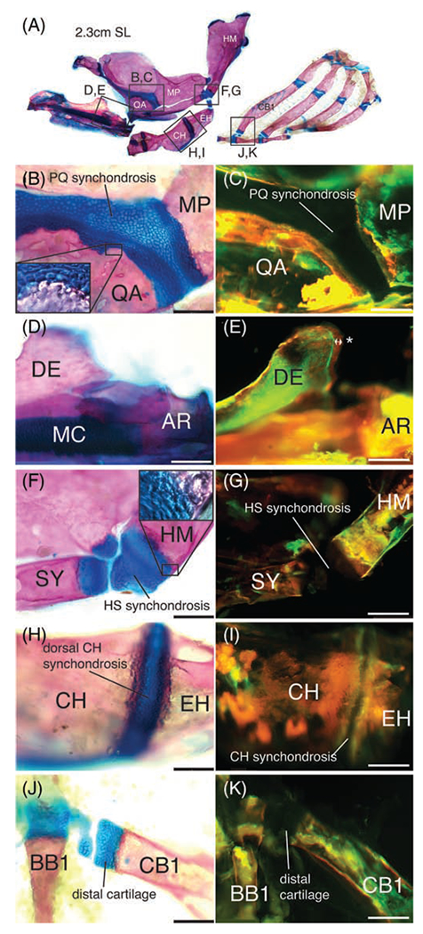
Endochondral ossification is a major bone forming mechanism in vertebrates, defects in which can result in skeletal dysplasia or craniofacial anomalies in humans. The zebrafish holds great potential to advance our understanding of endochondral growth zone development and genetics, yet several important aspects of its biology remain unexplored. Here we provide a comprehensive description of endochondral growth zones in the pharyngeal skeleton, including their developmental progression, cellular activity, and adult fates.
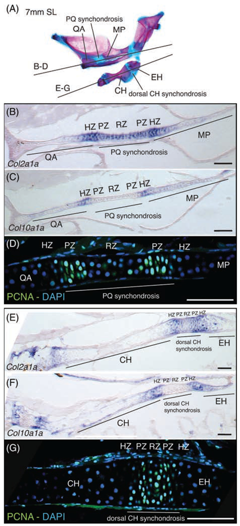
Postembryonic growth of the pharyngeal skeleton is supported by endochondral growth zones located either at skeletal epiphyses or synchondroses. Col2a1a and col10a1a in situ hybridization and anti-PCNA immunostaining identify resting-, hypertrophic- and proliferative zones, respectively, in pharyngeal synchondroses. Cellular hypertrophy and matrix deposition contribute little, if at all, to axial growth in most skeletal elements. Zebrafish endochondral growth zones develop during metamorphosis and arrest in adults.
Two endochondral growth zone configurations in the zebrafish pharyngeal skeleton produce either unidirectional (epiphyses) or bidirectional (synchondroses) growth. Cell proliferation drives endochondral growth and its modulation, in contrast to mammalian long bones in which bone length depends more on cell enlargement during hypertrophy and intramembranous ossification is the default mechanism of bone growth in zebrafish adults.
软骨内骨化是脊椎动物的主要成骨机制,其缺陷可导致人类骨骼发育不良或颅面异常。斑马鱼在推进我们对软骨内生长带发育和遗传学的理解方面具有巨大潜力,但它的生物学的几个重要方面仍未得到探索。 在这里,我们全面描述了咽颅骨骼中的软骨内生长带,包括其发育进程、细胞活性和成年后的命运。
咽颅骨骼的胚胎后生长由位于骨骼骺端或骺软骨处的软骨内生长带支持。 Col2a1a 和 col10a1a 原位杂交和抗 PCNA 免疫染色分别识别出咽骺软骨中的静止区、肥大区和增殖区。在大多数骨骼中,细胞肥大和基质沉积对轴向生长的贡献很小,如果有的话。 斑马鱼软骨内生长带在变态期间发育并在成年时停止。
斑马鱼咽颅骨骼中的两种软骨内生长带构型产生单向(骺端)或双向(骺软骨)生长。细胞增殖驱动软骨内生长及其调节,而与哺乳动物长骨不同,在哺乳动物长骨中,骨长度更多地取决于肥大期间细胞的增大,而软骨内成骨是成年斑马鱼骨生长的默认机制。