Department of Medical Biology, Amsterdam UMC, University of Amsterdam, Heart Center, Meibergdreef 9, P.O. Box 22660, 1100 DD Amsterdam, The Netherlands.
Department of Experimental Cardiology, Amsterdam UMC, University of Amsterdam, Heart Center, Meibergdreef 9, P.O. Box 22660, 1100 DD Amsterdam, The Netherlands.
Cardiovasc Res. 2021 Jul 27;117(9):2083-2091. doi: 10.1093/cvr/cvaa253.
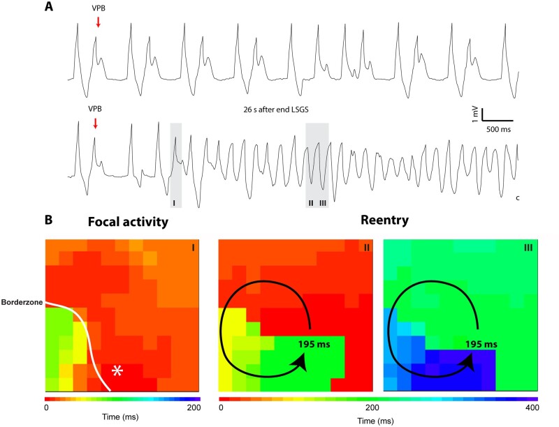
Enhanced sympathetic activity during acute ischaemia is arrhythmogenic, but the underlying mechanism is unknown. During ischaemia, a diastolic current flows from the ischaemic to the non-ischaemic myocardium. This 'injury' current can cause ventricular premature beats (VPBs) originating in the non-ischaemic myocardium, especially during a deeply negative T wave in the ischaemic zone. We reasoned that shortening of repolarization in myocardium adjacent to ischaemic myocardium increases the 'injury' current and causes earlier deeply negative T waves in the ischaemic zone, and re-excitation of the normal myocardium. We tested this hypothesis by activation and repolarization mapping during stimulation of the left stellate ganglion (LSG) during left anterior descending coronary artery (LAD) occlusion.
In nine pigs, five subsequent episodes of acute ischaemia, separated by 20 min of reperfusion, were produced by occlusion of the LAD and 121 epicardial local unipolar electrograms were recorded. During the third occlusion, left stellate ganglion stimulation (LSGS) was initiated after 3 min for a 30-s period, causing a shortening of repolarization in the normal myocardium by about 100 ms. This resulted in more negative T waves in the ischaemic zone and more VPBs than during the second, control, occlusion. Following the decentralization of the LSG (including removal of the right stellate ganglion and bilateral cervical vagotomy), fewer VPBs occurred during ischaemia without LSGS. During LSGS, the number of VPBs was similar to that recorded before decentralization.
LSGS, by virtue of shortening of repolarization in the non-ischaemic myocardium by about 100 ms, causes deeply negative T waves in the ischaemic tissue and VPBs originating from the normal tissue adjacent to the ischaemic border.
急性缺血期间增强的交感神经活动有致心律失常作用,但潜在机制尚不清楚。在缺血期间,从缺血区流向非缺血区的心肌存在舒张电流。这种“损伤”电流可导致起源于非缺血区的室性期前收缩(VPB),尤其是在缺血区深负 T 波时。我们推断,与缺血区相邻的心肌复极缩短会增加“损伤”电流,并导致缺血区更早出现深负 T 波,从而使正常心肌重新兴奋。我们通过在左前降支冠状动脉(LAD)闭塞期间刺激左侧星状神经节(LSG)时进行激活和复极标测来验证这一假设。
在 9 头猪中,通过 LAD 闭塞产生了五次急性缺血,每次缺血之间间隔 20 分钟再灌注,共记录了 121 个心外膜局部单极电图。在第三次闭塞期间,在 3 分钟后开始进行左侧星状神经节刺激(LSGS),持续 30 秒,导致正常心肌复极缩短约 100ms。这导致缺血区 T 波更加负向,VPB 比第二次对照闭塞时更多。在 LSG 去神经支配(包括去除右侧星状神经节和双侧颈迷走神经切断术)后,LSGS 期间缺血时发生的 VPB 减少,但 LSGS 期间发生的 VPB 数量与去神经支配前记录的相似。
LSGS 通过使非缺血区心肌复极缩短约 100ms,导致缺血组织中出现深负 T 波和起源于缺血边界附近正常组织的 VPB。