• 文献检索
  • 文档翻译
  • 深度研究
  • 学术资讯
  • Suppr Zotero 插件Zotero 插件
  • 邀请有礼
  • 套餐&价格
  • 历史记录
应用&插件
Suppr Zotero 插件Zotero 插件浏览器插件Mac 客户端Windows 客户端微信小程序
定价
高级版会员购买积分包购买API积分包
服务
文献检索文档翻译深度研究API 文档MCP 服务
关于我们
关于 Suppr公司介绍联系我们用户协议隐私条款
关注我们

Suppr 超能文献

核心技术专利:CN118964589B侵权必究
粤ICP备2023148730 号-1Suppr @ 2026

文献检索

告别复杂PubMed语法,用中文像聊天一样搜索,搜遍4000万医学文献。AI智能推荐,让科研检索更轻松。

立即免费搜索

文件翻译

保留排版,准确专业,支持PDF/Word/PPT等文件格式,支持 12+语言互译。

免费翻译文档

深度研究

AI帮你快速写综述,25分钟生成高质量综述,智能提取关键信息,辅助科研写作。

立即免费体验

青光眼小鼠模型中,CNS 轴突变性和视神经头部的运输缺陷先于视网膜神经节细胞的结构和功能丧失。

CNS axonal degeneration and transport deficits at the optic nerve head precede structural and functional loss of retinal ganglion cells in a mouse model of glaucoma.

机构信息

Department of Pharmacology and Neuroscience and the North Texas Eye Research Institute, IREB-535, University of North Texas Health Science Center, 3500 Camp Bowie Blvd, Fort Worth, TX, 76107, USA.

出版信息

Mol Neurodegener. 2020 Aug 27;15(1):48. doi: 10.1186/s13024-020-00400-9.

DOI:10.1186/s13024-020-00400-9
PMID:32854767
原文链接:https://pmc.ncbi.nlm.nih.gov/articles/PMC7457267/
Abstract

BACKGROUND

Glaucoma is a leading neurodegenerative disease affecting over 70 million individuals worldwide. Early pathological events of axonal degeneration and retinopathy in response to elevated intraocular pressure (IOP) are limited and not well-defined due to the lack of appropriate animal models that faithfully replicate all the phenotypes of primary open angle glaucoma (POAG), the most common form of glaucoma. Glucocorticoid (GC)-induced ocular hypertension (OHT) and its associated iatrogenic open-angle glaucoma share many features with POAG. Here, we characterized a novel mouse model of GC-induced OHT for glaucomatous neurodegeneration and further explored early pathological events of axonal degeneration in response to elevated IOP.

METHODS

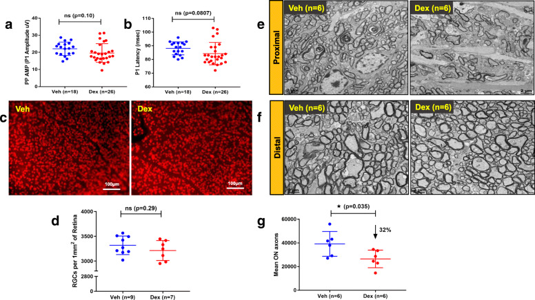
C57BL/6 J mice were periocularly injected with either vehicle or the potent GC, dexamethasone 21-acetate (Dex) once a week for 10 weeks. Glaucoma phenotypes including IOP, outflow facility, structural and functional loss of retinal ganglion cells (RGCs), optic nerve (ON) degeneration, gliosis, and anterograde axonal transport deficits were examined at various stages of OHT.

RESULTS

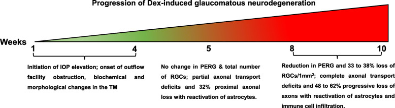
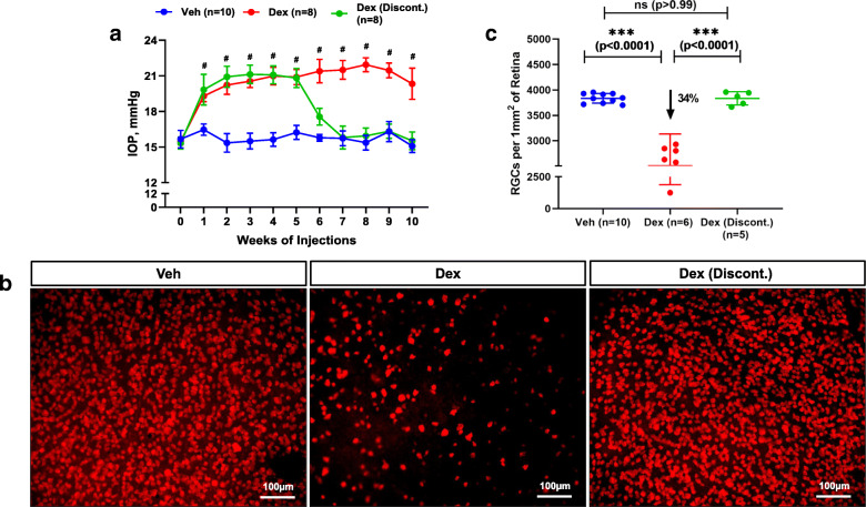
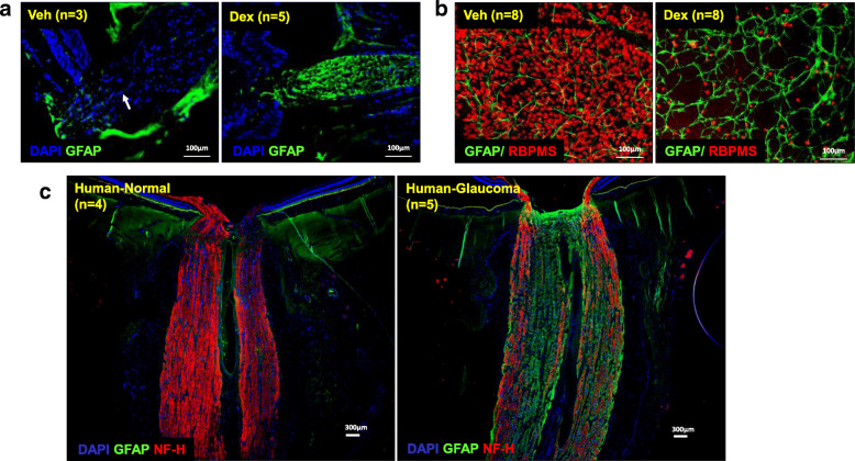
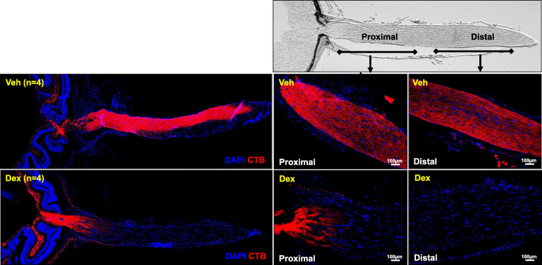
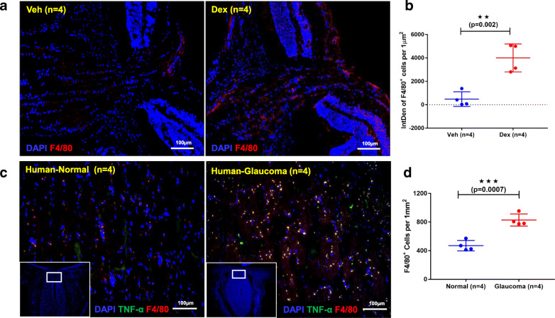
Prolonged treatment with Dex leads to glaucoma in mice similar to POAG patients including IOP elevation due to reduced outflow facility and dysfunction of trabecular meshwork, progressive ON degeneration and structural and functional loss of RGCs. Lowering of IOP rescued Dex-induced ON degeneration and RGC loss, suggesting that glaucomatous neurodegeneration is IOP dependent. Also, Dex-induced neurodegeneration was associated with activation of astrocytes, axonal transport deficits, ON demyelination, mitochondrial accumulation and immune cell infiltration in the optic nerve head (ONH) region. Our studies further show that ON degeneration precedes structural and functional loss of RGCs in Dex-treated mice. Axonal damage and transport deficits initiate at the ONH and progress toward the distal end of ON and target regions in the brain (i.e. superior colliculus). Most of anterograde transport was preserved during initial stages of axonal degeneration (30% loss) and complete transport deficits were only observed at the ONH during later stages of severe axonal degeneration (50% loss).

CONCLUSIONS

These findings indicate that ON degeneration and transport deficits at the ONH precede RGC structural and functional loss and provide a new potential therapeutic window for rescuing neuronal loss and restoring health of damaged axons in glaucoma.

摘要

背景

青光眼是一种主要的神经退行性疾病,影响着全球超过 7000 万人。由于缺乏能够真实复制原发性开角型青光眼(POAG)所有表型的合适动物模型,导致轴突变性和视网膜病变等早期病理事件受到限制,且尚未得到明确界定。POAG 是最常见的青光眼类型。糖皮质激素(GC)诱导的眼压升高(OHT)及其相关的医源性开角型青光眼与 POAG 有许多共同特征。在这里,我们描述了一种新的 GC 诱导的 OHT 小鼠模型,用于研究青光眼性神经退行性变,并进一步探讨了眼压升高时轴突变性的早期病理事件。

方法

每周一次向 C57BL/6J 小鼠眼周注射载体或强效 GC 地塞米松 21-醋酸酯(Dex),共 10 周。在 OHT 的不同阶段,检查眼压(IOP)、流出物通畅性、视网膜神经节细胞(RGC)的结构和功能丧失、视神经(ON)变性、神经胶质增生和顺行轴突运输缺陷等青光眼表型。

结果

Dex 的长期治疗会导致小鼠发生类似于 POAG 患者的青光眼,包括由于流出物通畅性降低和小梁网功能障碍导致的 IOP 升高、进行性 ON 变性以及 RGC 的结构和功能丧失。降低 IOP 可挽救 Dex 诱导的 ON 变性和 RGC 丧失,表明青光眼性神经退行性变与 IOP 有关。此外,Dex 诱导的神经退行性变与星形胶质细胞的激活、轴突运输缺陷、ON 脱髓鞘、线粒体积累和视神经头部(ONH)区域的免疫细胞浸润有关。我们的研究进一步表明,在 Dex 处理的小鼠中,ON 变性先于 RGC 的结构和功能丧失。轴突损伤和运输缺陷始于 ONH,并向 ON 的远端和大脑中的靶区(即上丘)进展。在轴突变性的初始阶段(丧失 30%),大部分顺行运输仍然保留,只有在严重轴突变性的后期阶段(丧失 50%)才会观察到 ONH 处的完全运输缺陷。

结论

这些发现表明,ONH 处的 ON 变性和运输缺陷先于 RGC 的结构和功能丧失,并为挽救神经元丧失和恢复青光眼受损轴突的健康提供了一个新的潜在治疗窗口。

https://cdn.ncbi.nlm.nih.gov/pmc/blobs/92b7/7457267/7a1cd4505a58/13024_2020_400_Fig12_HTML.jpg
https://cdn.ncbi.nlm.nih.gov/pmc/blobs/92b7/7457267/fde771cfa65d/13024_2020_400_Fig1_HTML.jpg
https://cdn.ncbi.nlm.nih.gov/pmc/blobs/92b7/7457267/ead3893934e7/13024_2020_400_Fig2_HTML.jpg
https://cdn.ncbi.nlm.nih.gov/pmc/blobs/92b7/7457267/f6fa19984b12/13024_2020_400_Fig3_HTML.jpg
https://cdn.ncbi.nlm.nih.gov/pmc/blobs/92b7/7457267/fd9c32481f39/13024_2020_400_Fig4_HTML.jpg
https://cdn.ncbi.nlm.nih.gov/pmc/blobs/92b7/7457267/4bb2dd465c83/13024_2020_400_Fig5_HTML.jpg
https://cdn.ncbi.nlm.nih.gov/pmc/blobs/92b7/7457267/ba3744a50555/13024_2020_400_Fig6_HTML.jpg
https://cdn.ncbi.nlm.nih.gov/pmc/blobs/92b7/7457267/89d944bb1887/13024_2020_400_Fig7_HTML.jpg
https://cdn.ncbi.nlm.nih.gov/pmc/blobs/92b7/7457267/5470204d7e9b/13024_2020_400_Fig8_HTML.jpg
https://cdn.ncbi.nlm.nih.gov/pmc/blobs/92b7/7457267/9cb3e6701000/13024_2020_400_Fig9_HTML.jpg
https://cdn.ncbi.nlm.nih.gov/pmc/blobs/92b7/7457267/8c62fb86b9f2/13024_2020_400_Fig10_HTML.jpg
https://cdn.ncbi.nlm.nih.gov/pmc/blobs/92b7/7457267/60d92f7be02d/13024_2020_400_Fig11_HTML.jpg
https://cdn.ncbi.nlm.nih.gov/pmc/blobs/92b7/7457267/7a1cd4505a58/13024_2020_400_Fig12_HTML.jpg

相似文献

1
CNS axonal degeneration and transport deficits at the optic nerve head precede structural and functional loss of retinal ganglion cells in a mouse model of glaucoma.青光眼小鼠模型中,CNS 轴突变性和视神经头部的运输缺陷先于视网膜神经节细胞的结构和功能丧失。
Mol Neurodegener. 2020 Aug 27;15(1):48. doi: 10.1186/s13024-020-00400-9.
2
Impaired axonal transport at the optic nerve head contributes to neurodegeneration in a novel Cre-inducible mouse model of myocilin glaucoma.在一种新型的肌纤蛋白青光眼Cre诱导小鼠模型中,视神经乳头处轴突运输受损导致神经退行性变。
bioRxiv. 2024 Nov 2:2024.09.18.613712. doi: 10.1101/2024.09.18.613712.
3
The optic nerve head is the site of axonal transport disruption, axonal cytoskeleton damage and putative axonal regeneration failure in a rat model of glaucoma.视盘是在青光眼大鼠模型中轴突运输中断、轴突细胞骨架损伤和可能的轴突再生失败的部位。
Acta Neuropathol. 2011 Jun;121(6):737-51. doi: 10.1007/s00401-011-0807-1. Epub 2011 Feb 11.
4
A method to quantify regional axonal transport blockade at the optic nerve head after short term intraocular pressure elevation in mice.一种在小鼠眼内压升高后定量视神经头区域轴突运输阻滞的方法。
Exp Eye Res. 2020 Jul;196:108035. doi: 10.1016/j.exer.2020.108035. Epub 2020 Apr 27.
5
The effects of age on mitochondria, axonal transport, and axonal degeneration after chronic IOP elevation using a murine ocular explant model.慢性眼压升高对小鼠眼组织体外模型中线粒体、轴突运输和轴突变性的影响。
Exp Eye Res. 2018 Jul;172:78-85. doi: 10.1016/j.exer.2018.04.001. Epub 2018 Apr 3.
6
A mouse ocular explant model that enables the study of living optic nerve head events after acute and chronic intraocular pressure elevation: Focusing on retinal ganglion cell axons and mitochondria.一种小鼠眼球外植体模型,可用于研究急性和慢性眼压升高后活体内视神经乳头的情况:聚焦于视网膜神经节细胞轴突和线粒体。
Exp Eye Res. 2017 Jul;160:106-115. doi: 10.1016/j.exer.2017.04.003. Epub 2017 Apr 14.
7
The dark phase intraocular pressure elevation and retinal ganglion cell degeneration in a rat model of experimental glaucoma.实验性青光眼大鼠模型中的暗相眼压升高和视网膜神经节细胞变性。
Exp Eye Res. 2013 Jul;112:21-8. doi: 10.1016/j.exer.2013.04.008. Epub 2013 Apr 18.
8
Assessment of retinal ganglion cell damage in glaucomatous optic neuropathy: Axon transport, injury and soma loss.青光眼性视神经病变中视网膜神经节细胞损伤的评估:轴突运输、损伤和胞体丢失。
Exp Eye Res. 2015 Dec;141:111-24. doi: 10.1016/j.exer.2015.06.006. Epub 2015 Jun 9.
9
Expression and activation of mitogen-activated protein kinases in the optic nerve head in a rat model of ocular hypertension.在眼高压大鼠模型中,视神经头部丝裂原活化蛋白激酶的表达和激活。
Mol Cell Neurosci. 2018 Apr;88:270-291. doi: 10.1016/j.mcn.2018.01.002. Epub 2018 Mar 20.
10
Retinal ganglion cell loss in a rat ocular hypertension model is sectorial and involves early optic nerve axon loss.大鼠眼高压模型中的视网膜神经节细胞丢失呈扇区性,且涉及早期视神经轴突丢失。
Invest Ophthalmol Vis Sci. 2011 Jan 21;52(1):434-41. doi: 10.1167/iovs.10-5856. Print 2011 Jan.

引用本文的文献

1
Modeling human retinal ganglion cell axonal outgrowth, development, and pathology using pluripotent stem cell-based microfluidic platforms.使用基于多能干细胞的微流控平台模拟人类视网膜神经节细胞轴突生长、发育及病理学过程。
bioRxiv. 2025 May 8:2025.05.02.651934. doi: 10.1101/2025.05.02.651934.
2
Maraviroc Prevents Optic Nerve Injury-Induced Retinal Ganglion Cell Apoptosis by Modulating the CCL5/CCR5/CTSS Axis.马拉维若通过调节CCL5/CCR5/CTSS轴预防视神经损伤诱导的视网膜神经节细胞凋亡。
Invest Ophthalmol Vis Sci. 2025 Jun 2;66(6):77. doi: 10.1167/iovs.66.6.77.
3
Protein misfolding and mitochondrial dysfunction in glaucoma.

本文引用的文献

1
Silicone oil-induced ocular hypertension and glaucomatous neurodegeneration in mouse.硅酮油诱导的小鼠眼内高压和青光眼性神经退行性变。
Elife. 2019 May 15;8:e45881. doi: 10.7554/eLife.45881.
2
Prior Exposure to Immunosuppressors Sensitizes Retinal Microglia and Accelerates Optic Nerve Regeneration in Zebrafish.先前暴露于免疫抑制剂会使视网膜小胶质细胞致敏,并加速斑马鱼视神经再生。
Mediators Inflamm. 2019 Feb 10;2019:6135795. doi: 10.1155/2019/6135795. eCollection 2019.
3
Inhibition of monocyte-like cell extravasation protects from neurodegeneration in DBA/2J glaucoma.
青光眼患者的蛋白质错误折叠与线粒体功能障碍
Front Cell Dev Biol. 2025 Apr 25;13:1595121. doi: 10.3389/fcell.2025.1595121. eCollection 2025.
4
SARM1: The Checkpoint of Axonal Degeneration in the Nervous System Disorders.SARM1:神经系统疾病中轴突退化的检查点。
Mol Neurobiol. 2025 Mar 17. doi: 10.1007/s12035-025-04835-3.
5
C/EBP Homologous Protein Expression in Retinal Ganglion Cells Induces Neurodegeneration in Mice.视网膜神经节细胞中C/EBP同源蛋白的表达诱导小鼠神经退行性变。
Int J Mol Sci. 2025 Feb 21;26(5):1858. doi: 10.3390/ijms26051858.
6
Damage and repair in retinal degenerative diseases: Molecular basis through clinical translation.视网膜退行性疾病中的损伤与修复:从分子基础到临床转化
Neural Regen Res. 2026 Apr 1;21(4):1383-1395. doi: 10.4103/NRR.NRR-D-24-01016. Epub 2025 Feb 24.
7
Impaired axonal transport contributes to neurodegeneration in a Cre-inducible mouse model of myocilin-associated glaucoma.在一种可诱导Cre的与肌纤蛋白相关青光眼小鼠模型中,轴突运输受损会导致神经退行性变。
JCI Insight. 2025 Jan 21;10(5):e188710. doi: 10.1172/jci.insight.188710.
8
Glaucomatous retinal ganglion cells: death and protection.青光眼性视网膜神经节细胞:死亡与保护
Int J Ophthalmol. 2025 Jan 18;18(1):160-167. doi: 10.18240/ijo.2025.01.20. eCollection 2025.
9
The role of RGC degeneration in the pathogenesis of glaucoma.视网膜神经节细胞变性在青光眼发病机制中的作用。
Int J Biol Sci. 2025 Jan 1;21(1):211-232. doi: 10.7150/ijbs.103222. eCollection 2025.
10
Identification of Sequential Molecular Mechanisms and Key Biomarkers in Early Glaucoma by Integrated Bioinformatics Analysis.通过综合生物信息学分析鉴定早期青光眼的序贯分子机制和关键生物标志物
Mol Neurobiol. 2025 Apr;62(4):4952-4970. doi: 10.1007/s12035-024-04563-0. Epub 2024 Nov 4.
抑制单核样细胞渗出可防止 DBA/2J 青光眼的神经退行性变。
Mol Neurodegener. 2019 Jan 22;14(1):6. doi: 10.1186/s13024-018-0303-3.
4
In vivo measurement of trabecular meshwork stiffness in a corticosteroid-induced ocular hypertensive mouse model.在皮质类固醇诱导的高眼压小鼠模型中测量小梁网硬度的体内测量。
Proc Natl Acad Sci U S A. 2019 Jan 29;116(5):1714-1722. doi: 10.1073/pnas.1814889116. Epub 2019 Jan 16.
5
Transforming growth factor β2 (TGFβ2) signaling plays a key role in glucocorticoid-induced ocular hypertension.转化生长因子 β2 (TGFβ2) 信号在糖皮质激素诱导的眼压升高中起着关键作用。
J Biol Chem. 2018 Jun 22;293(25):9854-9868. doi: 10.1074/jbc.RA118.002540. Epub 2018 May 9.
6
Axogenic mechanism enhances retinal ganglion cell excitability during early progression in glaucoma.轴突发生机制增强青光眼早期进展过程中视网膜神经节细胞的兴奋性。
Proc Natl Acad Sci U S A. 2018 Mar 6;115(10):E2393-E2402. doi: 10.1073/pnas.1714888115. Epub 2018 Feb 20.
7
Methods for Analyzing Endoplasmic Reticulum Stress in the Trabecular Meshwork of Glaucoma Models.青光眼模型小梁网内质网应激的分析方法
Methods Mol Biol. 2018;1695:121-134. doi: 10.1007/978-1-4939-7407-8_12.
8
ImageJ2: ImageJ for the next generation of scientific image data.ImageJ2:面向下一代科学图像数据的ImageJ。
BMC Bioinformatics. 2017 Nov 29;18(1):529. doi: 10.1186/s12859-017-1934-z.
9
Increased synthesis and deposition of extracellular matrix proteins leads to endoplasmic reticulum stress in the trabecular meshwork.细胞外基质蛋白的合成和沉积增加导致小梁网内质网应激。
Sci Rep. 2017 Nov 2;7(1):14951. doi: 10.1038/s41598-017-14938-0.
10
Early immune responses are independent of RGC dysfunction in glaucoma with complement component C3 being protective.早期免疫反应独立于青光眼视网膜神经节细胞功能障碍,补体成分C3具有保护作用。
Proc Natl Acad Sci U S A. 2017 May 9;114(19):E3839-E3848. doi: 10.1073/pnas.1608769114. Epub 2017 Apr 26.