USC Stevens Neuroimaging and Informatics Institute, Keck School of Medicine, University of Southern California, 2025 Zonal Ave., Los Angeles 90033, CA, USA.
USC Stevens Neuroimaging and Informatics Institute, Keck School of Medicine, University of Southern California, 2025 Zonal Ave., Los Angeles 90033, CA, USA; Department of Anatomy and Neurobiology, School of Basic Medical Science, Cheeloo College of Medicine, Shandong University, Jinan, Shandong, China.
Neuroimage. 2020 Dec;223:117301. doi: 10.1016/j.neuroimage.2020.117301. Epub 2020 Aug 28.
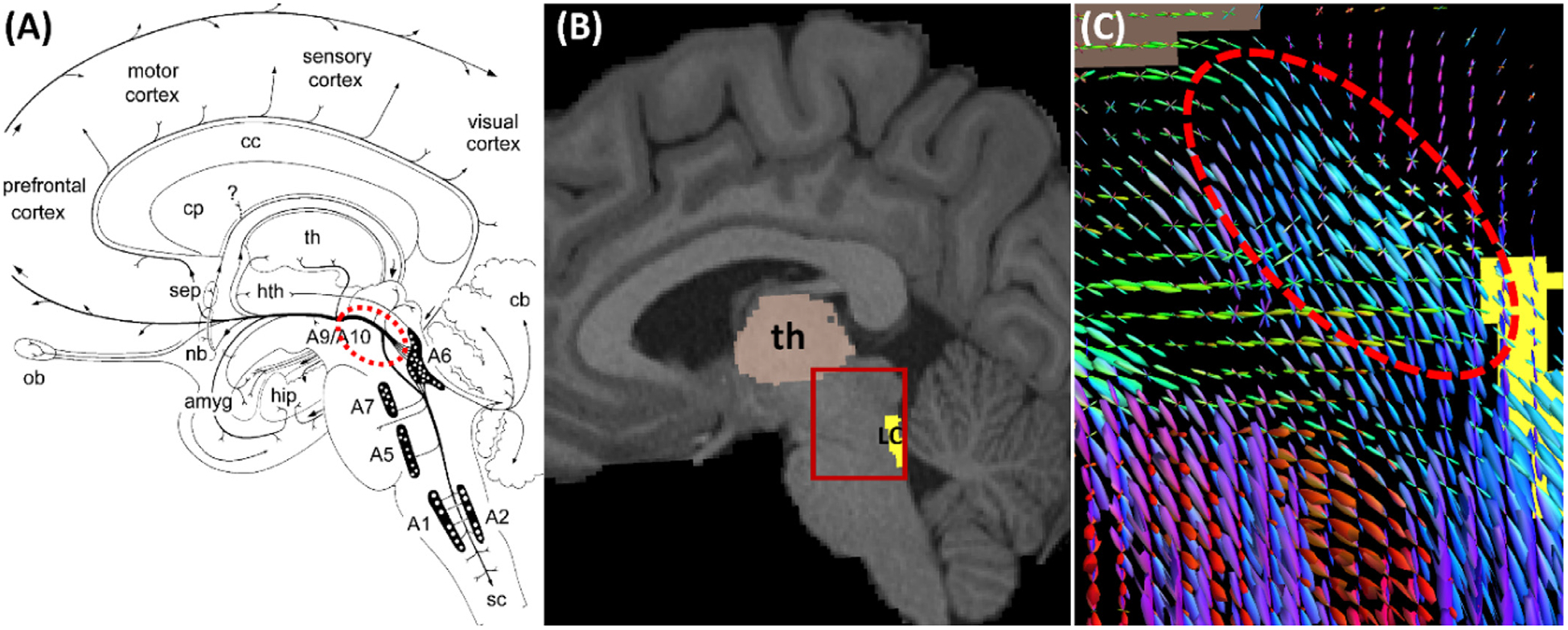
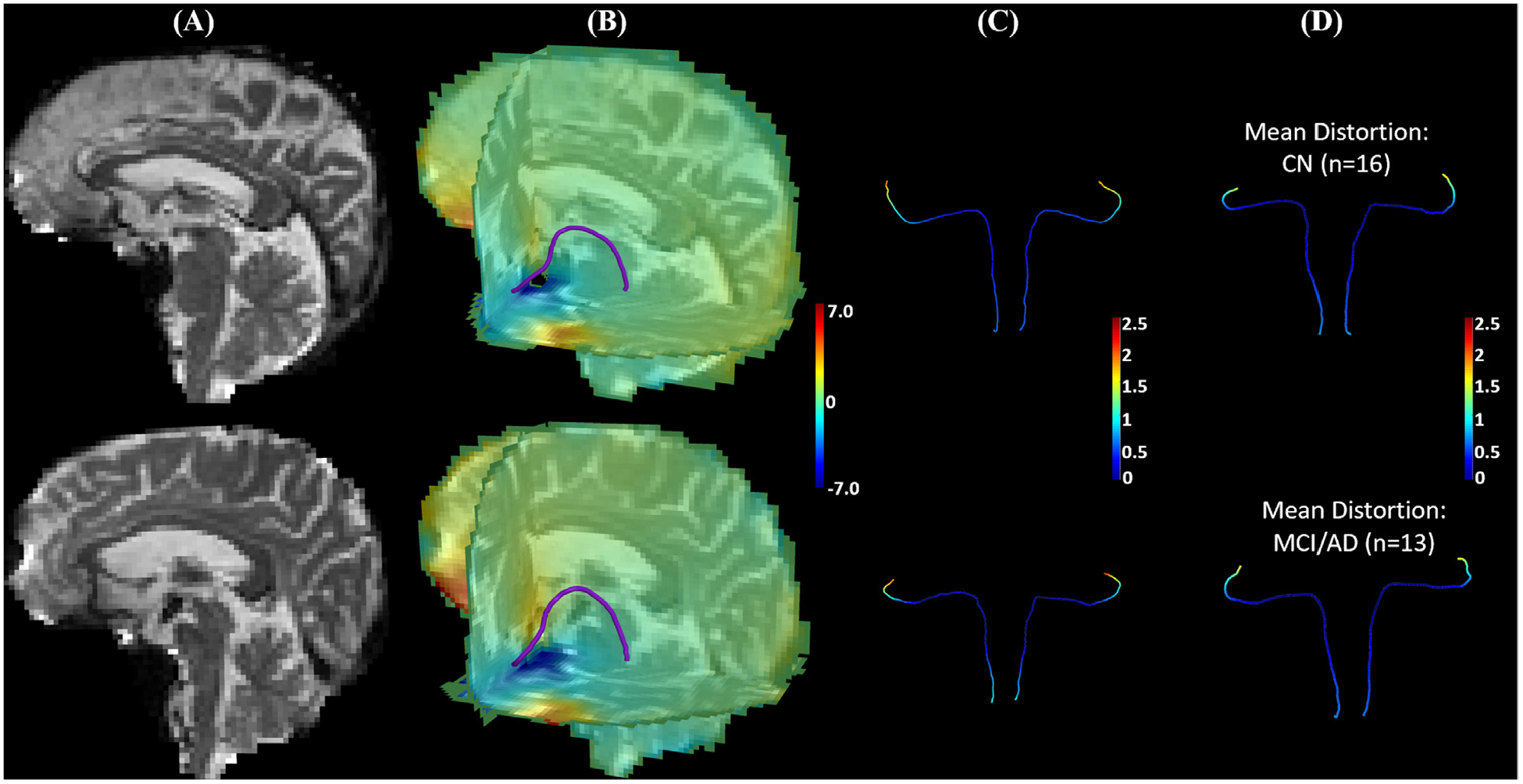
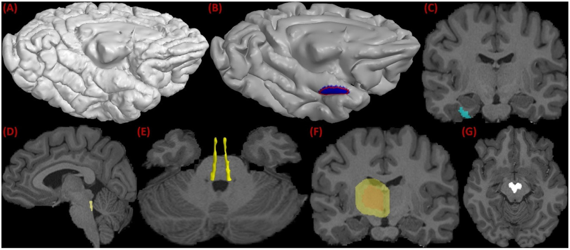
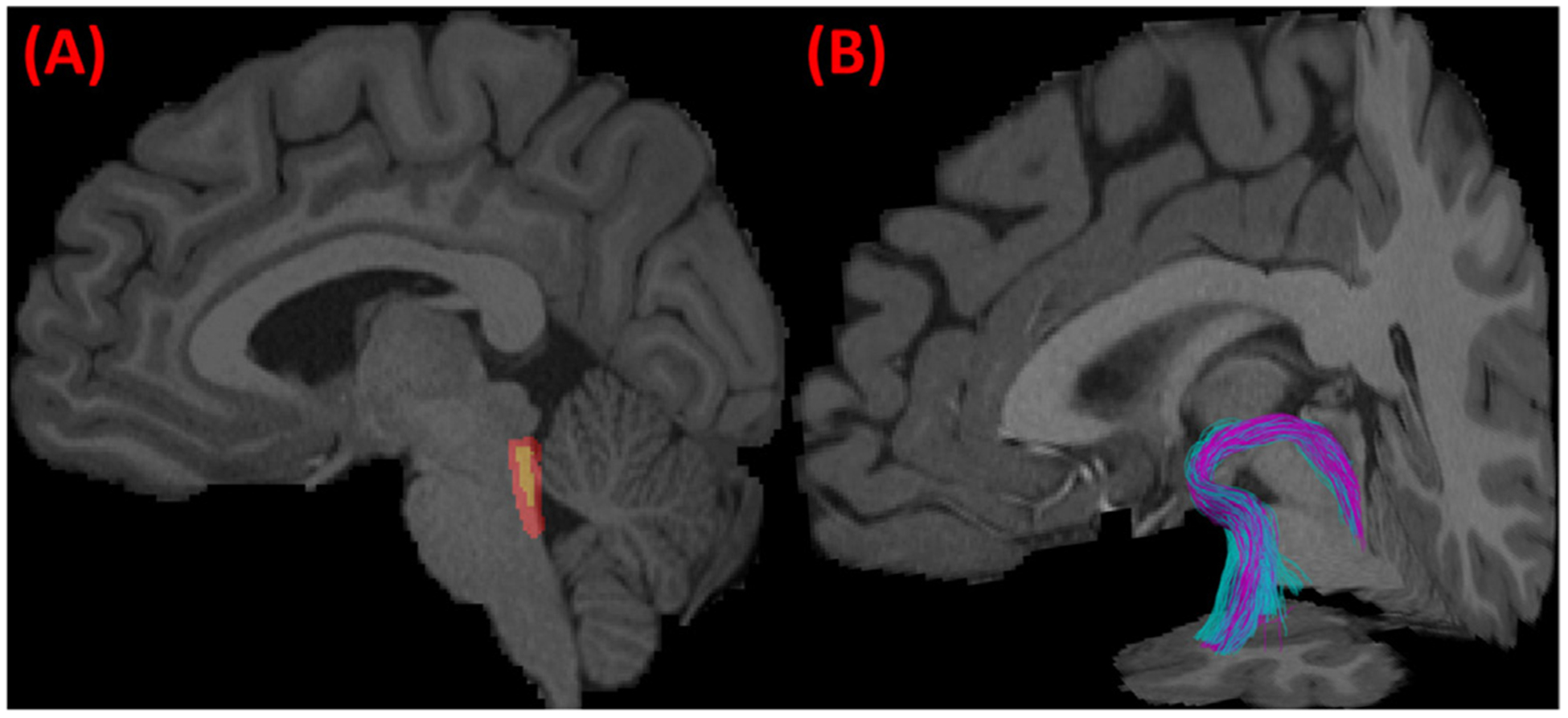
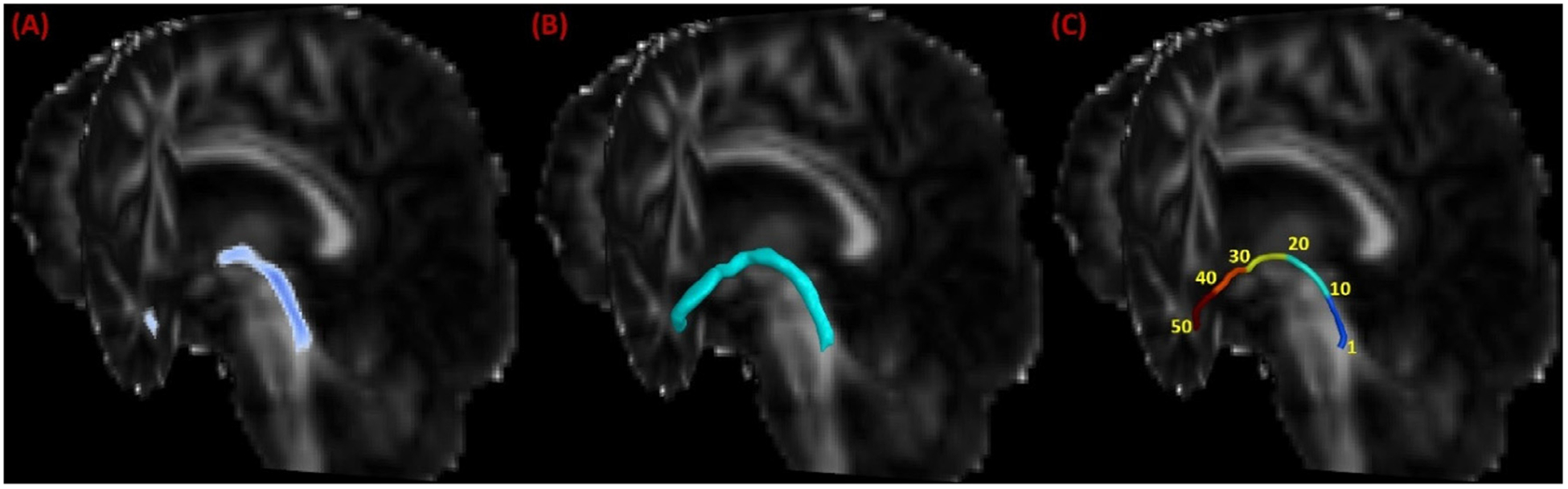
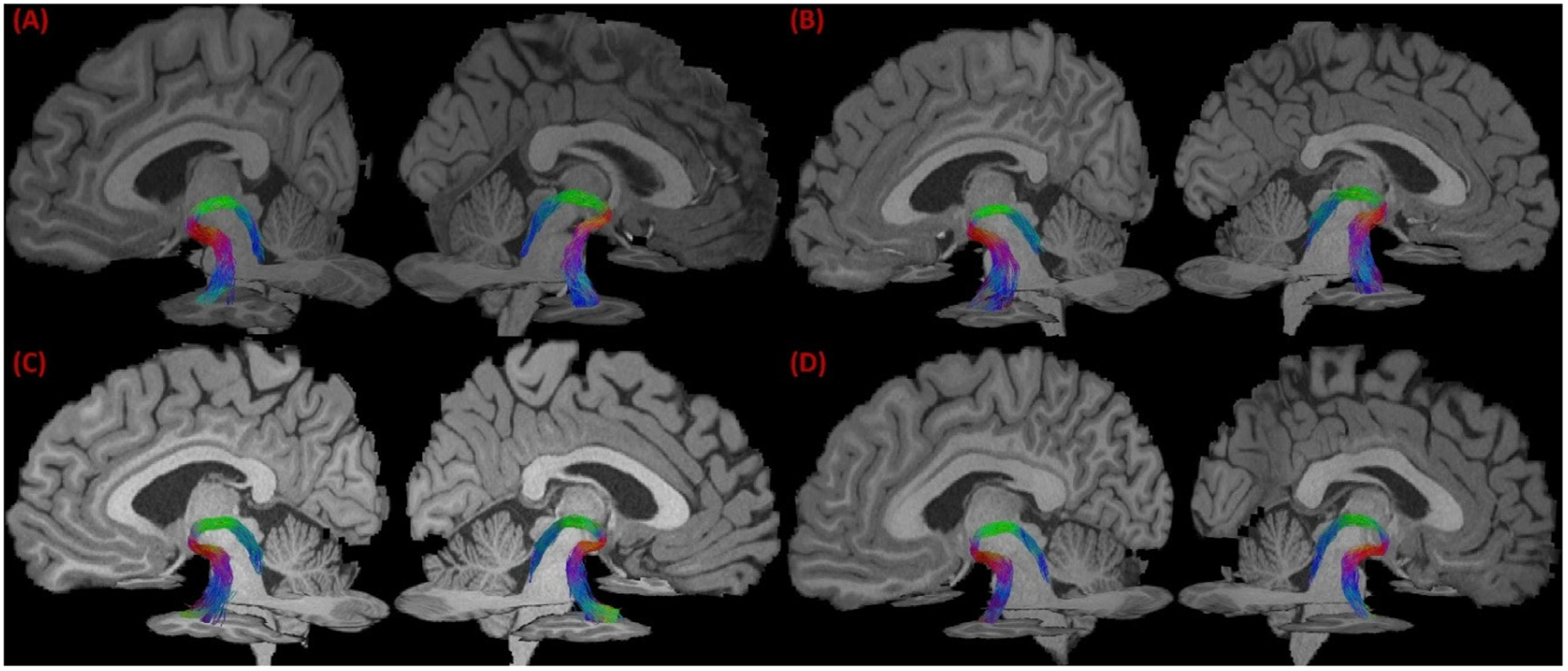
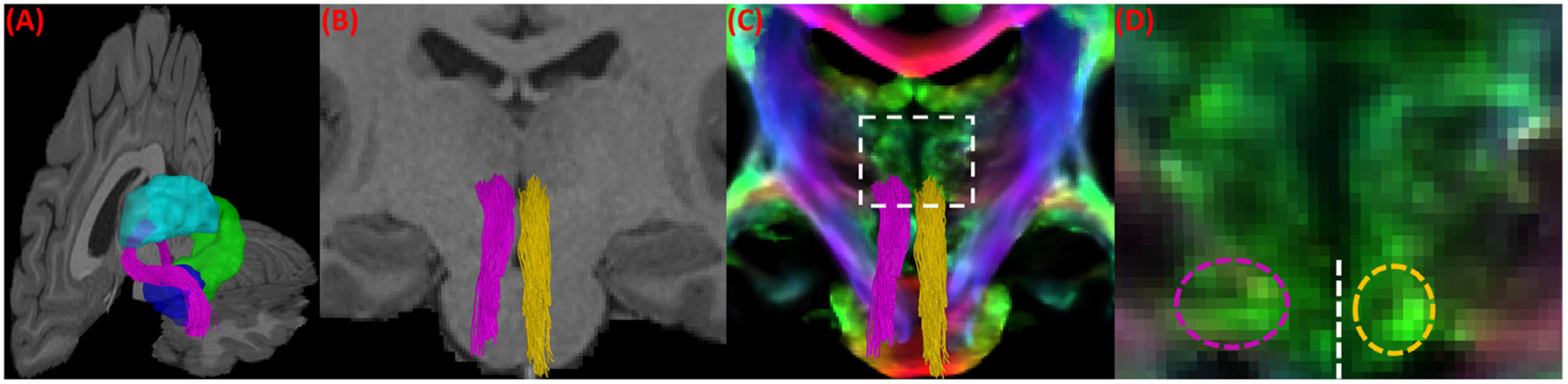
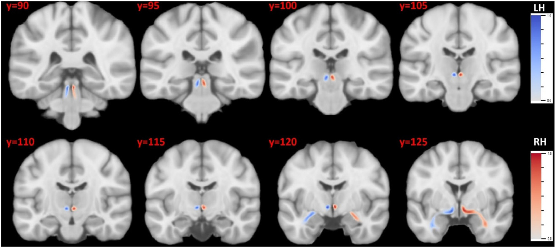
According to the latest Braak staging of Alzheimer's disease (AD), tau pathology occurs earliest in the brain in the locus coeruleus (LC) of the brainstem, then propagates to the transentorhinal cortex (TEC), and later to other neocortical regions. Recent animal and in vivo human brain imaging research also support the trans-axonal propagation of tau pathology. In addition, neurochemical studies link norepinephrine to behavioral symptoms in AD. It is thus critical to examine the integrity of the LC-TEC pathway in studying the early development of the disease, but there has been limited work in this direction. By leveraging the high-resolution and multi-shell diffusion MRI data from the Human Connectome Project (HCP), in this work we develop a novel method for the reconstruction of the LC-TEC pathway in a cohort of 40 HCP subjects carefully selected based on rigorous quality control of the residual distortion artifacts in the brainstem. A probabilistic atlas of the LC-TEC pathway of both hemispheres is then developed in the MNI152 space and distributed publicly on the NITRC website. To apply our atlas on clinical imaging data, we develop an automated approach to calculate the medial core of the LC-TEC pathway for localized analysis of connectivity changes. In a cohort of 138 subjects from the Alzheimer's Disease Neuroimaging Initiative (ADNI), we demonstrate the detection of the decreased fiber integrity in the LC-TEC pathways with increasing disease severity.
根据阿尔茨海默病(AD)的最新 Braak 分期,tau 病理学最早发生在脑干蓝斑(LC),然后传播到颞叶前脑(TEC),再传播到其他新皮质区域。最近的动物和体内人脑成像研究也支持 tau 病理学的跨轴突传播。此外,神经化学研究将去甲肾上腺素与 AD 的行为症状联系起来。因此,在研究疾病的早期发展时,检查 LC-TEC 通路的完整性至关重要,但在这方面的工作有限。通过利用人类连接组计划(HCP)的高分辨率和多壳扩散 MRI 数据,在这项工作中,我们开发了一种新方法,用于在一组 40 名 HCP 受试者中重建 LC-TEC 通路,这些受试者是根据脑干残余失真伪影的严格质量控制精心选择的。然后在 MNI152 空间中开发了双侧 LC-TEC 通路的概率图谱,并在 NITRC 网站上公开发布。为了将我们的图谱应用于临床成像数据,我们开发了一种自动方法来计算 LC-TEC 通路的内侧核心,以进行连接变化的局部分析。在来自阿尔茨海默病神经影像学倡议(ADNI)的 138 名受试者队列中,我们证明了随着疾病严重程度的增加,LC-TEC 通路的纤维完整性降低。