Xin Xiaonan, Zhu Yueyu, Xi Ruijie, Hao Yuhua
Department of Ophthalmology, Second Hospital of Hebei Medical University, Shijiazhuang 050000, China.
Saudi J Biol Sci. 2020 Sep;27(9):2491-2497. doi: 10.1016/j.sjbs.2020.06.039. Epub 2020 Jun 27.
To study the therapeutic effect and mechanism of levotinib on choroidal neovascularization (CNV) in mice.
45 healthy C57BL/6 mice were selected and randomly divided into three groups: control group (group A), model group (group B) and levotinib group (group C). The model of CNV in mice was established. The fluorescence leakage of choroidal lesions in mice was observed by fundus fluorescein angiography. The morphological changes of retinal vessels in mice were observed by retinal slice preparation, the pathological changes of eyeball tissues in mice were observed by hematoxylin-eosin (HE) staining, the expression of vascular endothelial growth factor (VEGF) in mice retina was detected by real-time quantitative fluorescence PCR, and the protein expression of VEGF in mice retina was detected by Western blotting.
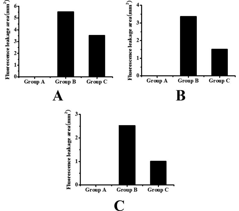
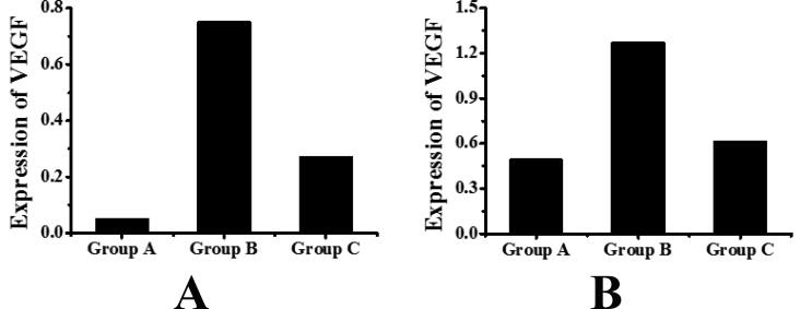
On the 7th, 14th and 21st day after modeling, compared with group B, the fluorescence leakage area of group C mice was significantly reduced, and the difference was statistically significant (P < 0.05). The morphology of retinal vessels in group A was normal. In group B, the retinal vessels showed large areas of ischemia without perfusion and abundant neovascularization clusters and capillaries. Compared with group B, the morphology of retinal vessels in group C was significantly improved. Group A mice had normal eyeball structure, group B mice had visible spindle-like damage to the inner and outer retina, while group C mice had significantly less spindle-like damage than group B. Compared with group A, group B mice had significantly higher expression of retinal VEGF and the difference was statistically significant (P < 0.05), but compared with group B mice, the expression of VEGF in the retina of mice in group C was significantly decreased, and the difference was statistically significant (P < 0.05). Compared with group A, the expression of VEGF in retina of group B mice was significantly increased, and the difference was statistically significant (P < 0.05). Compared with group B, the expression of VEGF in retina of group C mice was significantly decreased, and the difference was statistically significant (P < 0.05).
Levatinib has obvious therapeutic effect on CNV, which may be achieved by inhibiting the high expression of VEGF in CNV.
研究乐伐替尼对小鼠脉络膜新生血管(CNV)的治疗作用及机制。
选取45只健康C57BL/6小鼠,随机分为三组:对照组(A组)、模型组(B组)和乐伐替尼组(C组)。建立小鼠CNV模型。通过眼底荧光血管造影观察小鼠脉络膜病变的荧光渗漏情况。通过视网膜切片制备观察小鼠视网膜血管的形态变化,通过苏木精-伊红(HE)染色观察小鼠眼球组织的病理变化,通过实时定量荧光PCR检测小鼠视网膜中血管内皮生长因子(VEGF)的表达,通过蛋白质免疫印迹法检测小鼠视网膜中VEGF的蛋白表达。
建模后第7天、14天和21天,与B组相比,C组小鼠的荧光渗漏面积显著减小,差异有统计学意义(P<0.05)。A组视网膜血管形态正常。B组视网膜血管出现大片缺血无灌注区,并有丰富的新生血管簇和毛细血管。与B组相比,C组视网膜血管形态明显改善。A组小鼠眼球结构正常,B组小鼠视网膜内、外可见梭形损伤,而C组小鼠的梭形损伤明显少于B组。与A组相比,B组小鼠视网膜VEGF表达显著升高,差异有统计学意义(P<0.05),但与B组小鼠相比,C组小鼠视网膜VEGF表达显著降低,差异有统计学意义(P<0.05)。与A组相比,B组小鼠视网膜VEGF表达显著升高,差异有统计学意义(P<0.05)。与B组相比,C组小鼠视网膜VEGF表达显著降低,差异有统计学意义(P<0.05)。
乐伐替尼对CNV有明显治疗作用,其机制可能是通过抑制CNV中VEGF的高表达实现的。