Jiemy William F, van Sleen Yannick, van der Geest Kornelis Sm, Ten Berge Hilde A, Abdulahad Wayel H, Sandovici Maria, Boots Annemieke Mh, Heeringa Peter, Brouwer Elisabeth
Department of Pathology and Medical Biology University of Groningen University Medical Center Groningen Groningen The Netherlands.
Faculty of Applied Science UCSI University UCSI Heights Cheras, Kuala Lumpur Malaysia.
Clin Transl Immunology. 2020 Aug 27;9(9):e1164. doi: 10.1002/cti2.1164. eCollection 2020.
To determine the presence and spatial distribution of different macrophage phenotypes, governed by granulocyte macrophage colony-stimulating factor (GM-CSF) and macrophage colony-stimulating factor (M-CSF) skewing signals, in giant cell arteritis (GCA) lesions.
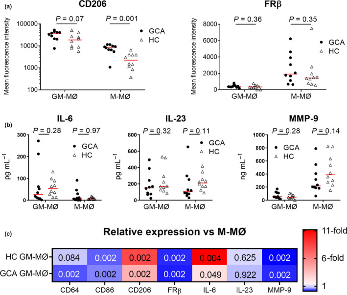
Temporal artery biopsies (TABs, = 11) from treatment-naive GCA patients, aorta samples from GCA-related aneurysms ( = 10) and atherosclerosis ( = 10) were stained by immunohistochemistry targeting selected macrophage phenotypic markers, cytokines, matrix metalloproteinases (MMPs) and growth factors. macrophage differentiation ( = 10) followed by flow cytometry, Luminex assay and ELISA were performed to assess whether GM-CSF and M-CSF are drivers of macrophage phenotypic heterogeneity.
A distinct spatial distribution pattern of macrophage phenotypes in TABs was identified. CD206/MMP-9 macrophages were located at the site of tissue destruction, whereas FRβ macrophages were located in the inner intima of arteries with high degrees of intimal hyperplasia. Notably, this pattern was also observed in macrophage-rich areas in GCA aortas but not in atherosclerotic aortas. Flow cytometry showed that GM-CSF treatment highly upregulated CD206 expression, while FRβ was expressed by M-CSF-skewed macrophages, only. Furthermore, localised expression of GM-CSF and M-CSF was detected, likely contributing to macrophage heterogeneity in the vascular wall.
Our data document a distinct spatial distribution pattern of CD206/MMP-9 macrophages and FRβ macrophages in GCA linked to tissue destruction and intimal proliferation, respectively. We suggest that these distinct macrophage phenotypes are skewed by sequential GM-CSF and M-CSF signals. Our study adds to a better understanding of the development and functional role of macrophage phenotypes in the pathogenesis of GCA and opens opportunities for the design of macrophage-targeted therapies.
确定由粒细胞巨噬细胞集落刺激因子(GM-CSF)和巨噬细胞集落刺激因子(M-CSF)偏向信号调控的不同巨噬细胞表型在巨细胞动脉炎(GCA)病变中的存在情况及空间分布。
对未经治疗的GCA患者的颞动脉活检标本(n = 11)、GCA相关动脉瘤(n = 10)和动脉粥样硬化(n = 10)的主动脉样本进行免疫组织化学染色,以检测选定的巨噬细胞表型标志物、细胞因子、基质金属蛋白酶(MMPs)和生长因子。通过流式细胞术、Luminex检测和酶联免疫吸附测定(ELISA)对巨噬细胞分化(n = 10)进行检测,以评估GM-CSF和M-CSF是否为巨噬细胞表型异质性的驱动因素。
在颞动脉活检标本中确定了巨噬细胞表型的独特空间分布模式。CD206/MMP-9巨噬细胞位于组织破坏部位,而FRβ巨噬细胞位于内膜高度增生动脉的内膜内层。值得注意的是,在GCA主动脉富含巨噬细胞的区域也观察到这种模式,但在动脉粥样硬化主动脉中未观察到。流式细胞术显示,GM-CSF处理可高度上调CD206表达,而FRβ仅由M-CSF偏向的巨噬细胞表达。此外,检测到GM-CSF和M-CSF的局部表达,这可能导致血管壁中的巨噬细胞异质性。
我们的数据证明了GCA中CD206/MMP-9巨噬细胞和FRβ巨噬细胞的独特空间分布模式,分别与组织破坏和内膜增殖相关。我们认为这些不同的巨噬细胞表型由连续的GM-CSF和M-CSF信号偏向。我们的研究有助于更好地理解巨噬细胞表型在GCA发病机制中的发展和功能作用,并为设计巨噬细胞靶向治疗提供了机会。