Department of Electrical Engineering, National Taiwan University of Science and Technology, Taipei, Taiwan.
Department of Automatic Control Engineering, Feng Chia University, Taichung, Taiwan.
NMR Biomed. 2021 Jan;34(1):e4408. doi: 10.1002/nbm.4408. Epub 2020 Sep 4.
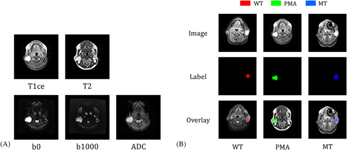
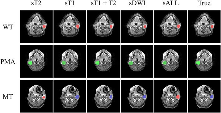
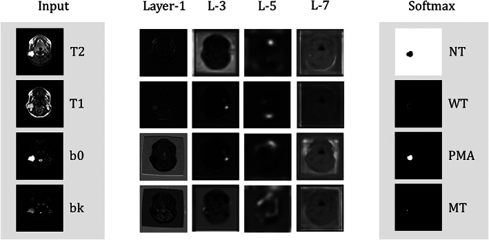
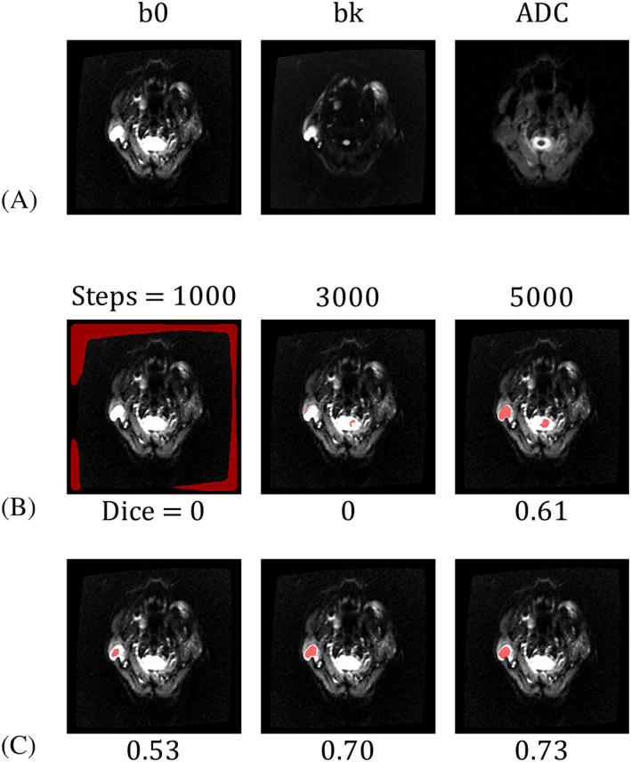
Various MRI sequences have shown their potential to discriminate parotid gland tumors, including but not limited to T -weighted, postcontrast T -weighted, and diffusion-weighted images. In this study, we present a fully automatic system for the diagnosis of parotid gland tumors by using deep learning methods trained on multimodal MRI images. We used a two-dimensional convolution neural network, U-Net, to segment and classify parotid gland tumors. The U-Net model was trained with transfer learning, and a specific design of the batch distribution optimized the model accuracy. We also selected five combinations of MRI contrasts as the input data of the neural network and compared the classification accuracy of parotid gland tumors. The results indicated that the deep learning model with diffusion-related parameters performed better than those with structural MR images. The performance results (n = 85) of the diffusion-based model were as follows: accuracy of 0.81, 0.76, and 0.71, sensitivity of 0.83, 0.63, and 0.33, and specificity of 0.80, 0.84, and 0.87 for Warthin tumors, pleomorphic adenomas, and malignant tumors, respectively. Combining diffusion-weighted and contrast-enhanced T -weighted images did not improve the prediction accuracy. In summary, the proposed deep learning model could classify Warthin tumor and pleomorphic adenoma tumor but not malignant tumor.
各种 MRI 序列已显示出其区分腮腺肿瘤的潜力,包括但不限于 T1 加权、增强后 T1 加权和弥散加权图像。在这项研究中,我们提出了一种基于深度学习方法的全自动系统,该系统可通过对多模态 MRI 图像进行训练来诊断腮腺肿瘤。我们使用二维卷积神经网络 U-Net 对腮腺肿瘤进行分割和分类。U-Net 模型通过迁移学习进行训练,批次分布的特定设计优化了模型的准确性。我们还选择了五种 MRI 对比组合作为神经网络的输入数据,并比较了腮腺肿瘤的分类准确性。结果表明,具有扩散相关参数的深度学习模型的性能优于具有结构 MRI 图像的模型。基于扩散的模型的性能结果(n=85)如下:Warthin 肿瘤、多形性腺瘤和恶性肿瘤的准确率分别为 0.81、0.76 和 0.71,敏感度分别为 0.83、0.63 和 0.33,特异性分别为 0.80、0.84 和 0.87。结合弥散加权和增强 T1 加权图像并没有提高预测的准确性。综上所述,所提出的深度学习模型可以对 Warthin 肿瘤和多形性腺瘤进行分类,但不能对恶性肿瘤进行分类。