Orthopaedic and Trauma Unit, Department of Basic Medical Sciences, Neuroscience and Sense Organs, School of Medicine, University of Bari "Aldo Moro", AOU Consorziale "Policlinico", Piazza Giulio Cesare 11, 70100, Bari, Italy.
Department of Orthopaedic and Trauma Surgery, Ospedali Riuniti, Ancona, Italy.
Aging Clin Exp Res. 2021 Jun;33(6):1627-1633. doi: 10.1007/s40520-020-01682-1. Epub 2020 Sep 9.
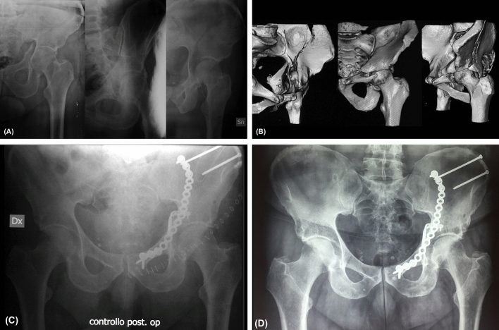
Osteoporotic acetabular fractures frequently involve the quadrilateral plate (QP), a flat and thin bone constituting the medial wall of the acetabulum. This study aims to assess the impact of the quality of osteoporotic QP fractures reduction on the patients' functional recovery, at 24 months follow-up.
Patients referring with osteoporotic QP fractures to our Level I trauma centre were prospectively recruited.
patients aged 60 years old or older; osteoporosis, defined as Dual-energy X-ray Absorptiometry (DXA) T-score ≤ - 2.5; acute acetabular fracture; anatomic or good fracture reduction according to Matta on postoperative CT.
moderate cognitive impairment (defined as Mini-Mental State Examination < 19); a history of malignant neoplasm; concomitant fractures in other sites; traumatic head injury; lower limb joint prostheses; patient not able to walk independently before trauma; poor fracture reduction, according to Matta, on postoperative CT. All the QP fractures were surgically managed. After surgery, the reduction of each QP fracture was classified as anatomical (displacement 0-1 mm), good (displacement 2-3 mm) and poor (displacement > 3 mm) on postoperative CT. Based on this classification: patients with a poor fracture reduction were excluded from this study, patients with an anatomical reduction were recruited in Group-A and patients with a good reduction in Group-B. All the patients underwent a clinical and radiographic 24-months follow-up.
68 patients (males 38; females 30; mean age 68.6 years old; range 60-79) were finally included in in the study. No cases of open fractures or concomitant pelvic ring fractures were observed. Based on the post-operative CT, 39 patients showed an anatomic fracture reduction (Group-A) while the remaining 29 patients revealed a good fracture reduction (Group-B). Complication rates and mean clinical scores showed no significant differences between groups, at 24-months follow-up.
In this study, the functional recovery at 24 months follow-up showed no significant differences in elderly patients with QP fracture undergoing anatomical reconstruction (displacement 0-1 mm) compared to patients receiving a good QP fracture reconstruction (displacement ≤ 3 mm).
骨质疏松性髋臼骨折常累及四边形板(QP),QP 是构成髋臼内侧壁的扁平薄骨。本研究旨在评估骨质疏松性 QP 骨折复位质量对患者功能恢复的影响,随访时间为 24 个月。
前瞻性招募到我们的 I 级创伤中心就诊的骨质疏松性 QP 骨折患者。
年龄 60 岁或以上;骨质疏松症,定义为双能 X 线吸收法(DXA)T 评分≤-2.5;急性髋臼骨折;根据术后 CT ,解剖复位或良好复位符合马塔标准。
中度认知障碍(定义为简易精神状态检查<19);恶性肿瘤病史;其他部位骨折;创伤性头部损伤;下肢关节假体;受伤前不能独立行走;术后 CT 显示复位不良,符合马塔标准。所有 QP 骨折均采用手术治疗。手术后,根据术后 CT 将每个 QP 骨折的复位分为解剖复位(移位 0-1mm)、良好复位(移位 2-3mm)和不良复位(移位>3mm)。基于此分类:复位不良的患者排除在本研究之外,解剖复位的患者纳入 A 组,良好复位的患者纳入 B 组。所有患者均接受了 24 个月的临床和影像学随访。
最终纳入 68 例患者(男 38 例,女 30 例;平均年龄 68.6 岁;60-79 岁)。未观察到开放性骨折或合并骨盆环骨折。根据术后 CT,39 例患者显示解剖复位(A 组),其余 29 例患者显示良好复位(B 组)。24 个月随访时,并发症发生率和平均临床评分在两组间无显著差异。
在本研究中,24 个月随访时,接受解剖复位(移位 0-1mm)的 QP 骨折老年患者与接受良好 QP 骨折复位(移位≤3mm)的患者相比,功能恢复无显著差异。