Aghashani Armeti, Verstraete Frank J M, Arzi Boaz
William R. Pritchard Veterinary Medical Teaching Hospital, School of Veterinary Medicine, University of California, Davis, Davis, CA, United States.
Department of Surgical and Radiological Sciences, School of Veterinary Medicine, University of California, Davis, Davis, CA, United States.
Front Vet Sci. 2020 Aug 13;7:482. doi: 10.3389/fvets.2020.00482. eCollection 2020.
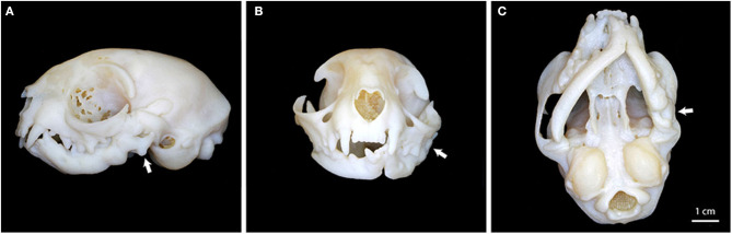
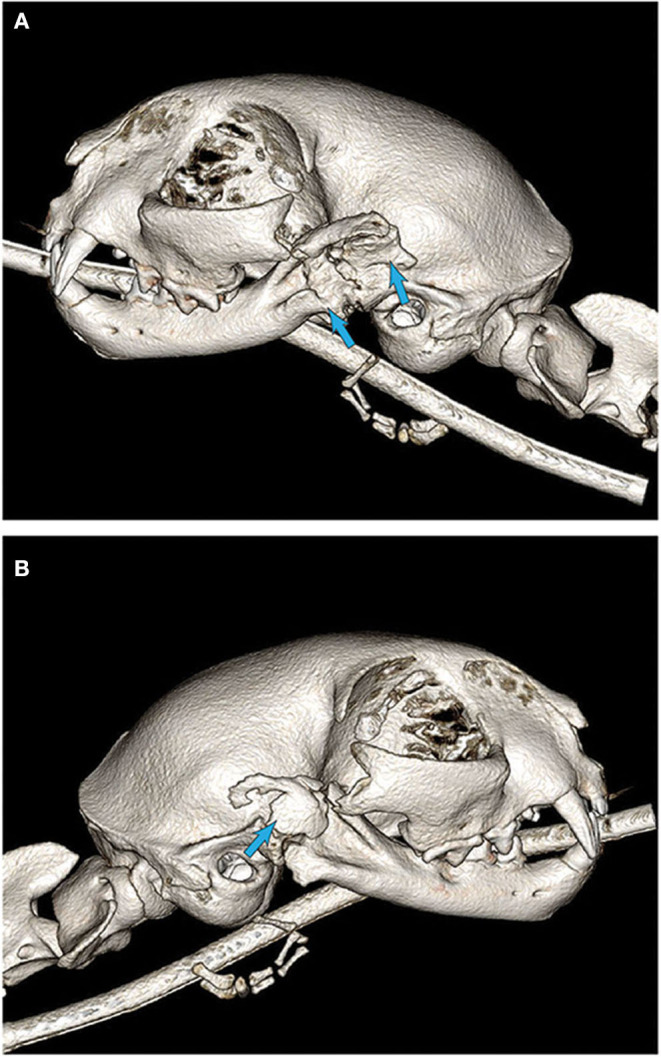
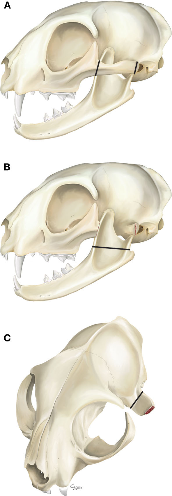
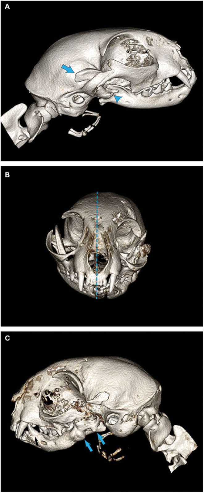
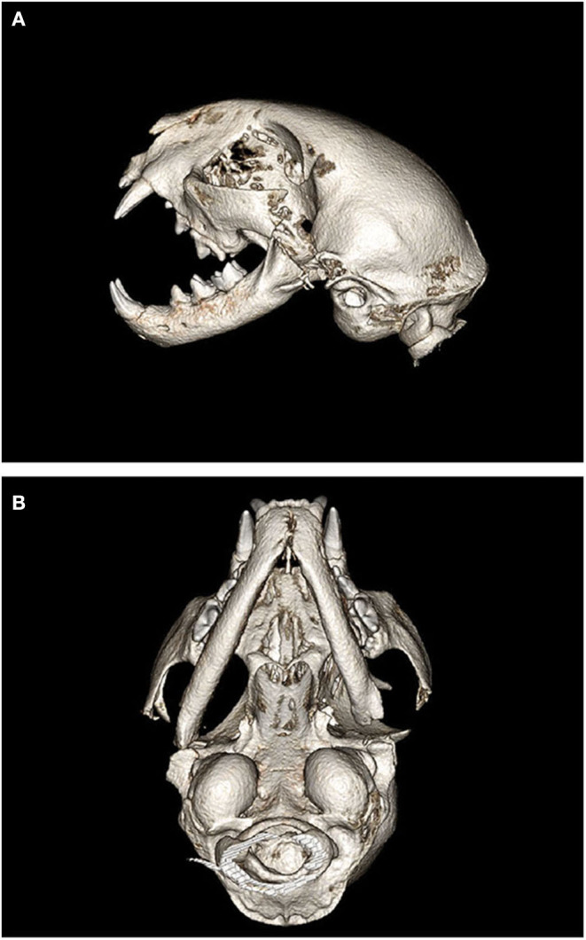
Temporomandibular joint (TMJ) ankylosis is defined as fibrous or bony fusion of the mandibular head of the condylar process and the mandibular fossa of the squamous part of the temporal bone. Ankylosis of the TMJ may be intraarticular, extraarticular, or both. The objective of this report is to describe the surgical planning, technique, and outcome of gap arthroplasty for extensive TMJ ankylosis in cats. Client-owned cats ( = 7) were examined clinically and surgical planning included the use of cone-beam computed tomography (CBCT) and tridimensional (3D) printed models. In six of the seven cats, temporary tracheostomy intubation was required. Gap arthroplasty included zygomectomy, coronoidectomy, condylectomy, as well as fossectomy (removal of the mandibular fossa of the temporal bone) and was performed using a piezosurgical unit. In all seven cats, gap arthroplasty was performed without surgical complications. In addition, a clinically acceptable mouth opening was achieved in all cases. However, a noticeable mandibular instability was observed. Medium-term follow-up demonstrated acceptable quality of life with one case of recurrence of ankylosis requiring repeated bilateral surgery, and a second case with recurrence of ankylosis not requiring surgical intervention at the time of manuscript preparation. We concluded that TMJ gap arthroplasty in cats is a salvage procedure indicated in cases of severe intraarticular and extraarticular ankylosis. Diagnostic imaging by means of CBCT and 3D printing are essential for precise surgical planning. The use of a piezosurgical unit allows for safe and precise ostectomy. Clinically, despite the resulting mandibular instability, appropriate prehension of food and water was possible.
颞下颌关节(TMJ)强直被定义为髁突下颌头与颞骨鳞部下颌窝之间的纤维性或骨性融合。颞下颌关节强直可能是关节内的、关节外的或两者皆有。本报告的目的是描述猫广泛颞下颌关节强直的间隙关节成形术的手术规划、技术和结果。对客户拥有的猫(n = 7)进行了临床检查,手术规划包括使用锥形束计算机断层扫描(CBCT)和三维(3D)打印模型。在7只猫中的6只中,需要进行临时气管切开插管。间隙关节成形术包括颧骨切除术、冠突切除术、髁突切除术以及颞骨下颌窝切除术(切除颞骨下颌窝),并使用压电手术装置进行。在所有7只猫中,间隙关节成形术均未出现手术并发症。此外,所有病例均实现了临床上可接受的开口度。然而,观察到明显的下颌骨不稳定。中期随访显示生活质量可接受,1例强直复发需要再次进行双侧手术,另1例强直复发在撰写本文时无需手术干预。我们得出结论,猫的颞下颌关节间隙关节成形术是一种挽救性手术,适用于严重的关节内和关节外强直病例。通过CBCT和3D打印进行诊断成像对于精确的手术规划至关重要。使用压电手术装置可实现安全精确的截骨术。临床上,尽管会导致下颌骨不稳定,但仍可适当摄取食物和水。