Marshall S Alex, McClain Justin A, Wooden Jessica I, Nixon Kimberly
Department of Pharmaceutical Sciences, University of Kentucky, Lexington, KY, United States.
Division of Pharmacology & Toxicology, College of Pharmacy, The University of Texas, Austin, TX, United States.
Front Neuroanat. 2020 Aug 13;14:52. doi: 10.3389/fnana.2020.00052. eCollection 2020.
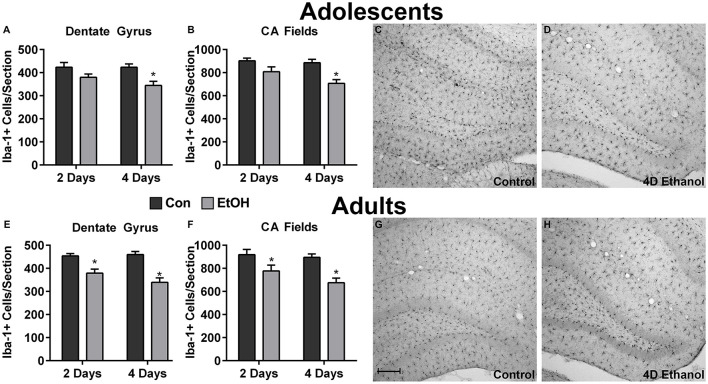
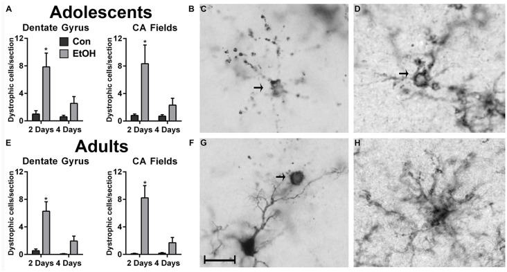
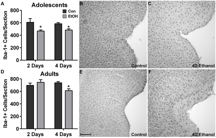
Microglia are dynamic cells that have roles in neuronal plasticity as well as in recovery responses following neuronal injury. Although many hypothesize that hyperactivation of microglia contributes to alcohol-induced neuropathology, in other neurodegenerative conditions disruption of normal microglial processes also contributes to neuronal loss, particularly as microglia become dystrophic or dysfunctional. Based on the observation of a striking, abnormal morphology in microglia during binge-like ethanol exposure, the present study investigated the impact of excessive ethanol exposure on microglia number and dystrophic morphology in a model of alcohol dependence that includes neurodegeneration in both adult and adolescent rats. Following 2- and 4-day binge ethanol exposure, the number of microglia was decreased in the hippocampus and the perirhinal and entorhinal cortices of both adult and adolescent rats. Furthermore, a significant number of microglia with a dystrophic morphology were observed in ethanol-exposed tissue, accompanied by a significant decrease in brain-derived neurotrophic factor (BDNF) expression in the hippocampus. Together these findings suggest another means by which microglia may contribute to alcohol-induced neurodegeneration, specifically dystrophic microglia and/or loss of microglia may disrupt homeostatic and recovery mechanisms. These results demonstrate that microglia also degenerate with excessive alcohol exposure, which has important implications for understanding the role of microglia-and specifically their contributions to plasticity and neuronal survival-in neurodegenerative disease.
小胶质细胞是动态细胞,在神经元可塑性以及神经元损伤后的恢复反应中发挥作用。尽管许多人推测小胶质细胞的过度激活会导致酒精诱导的神经病理学,但在其他神经退行性疾病中,正常小胶质细胞过程的破坏也会导致神经元丢失,特别是当小胶质细胞变得营养不良或功能失调时。基于在类似暴饮的乙醇暴露期间观察到小胶质细胞中显著的异常形态,本研究在一个酒精依赖模型中研究了过量乙醇暴露对小胶质细胞数量和营养不良形态的影响,该模型包括成年和青少年大鼠的神经退行性变。在进行2天和4天的暴饮乙醇暴露后,成年和青少年大鼠海马体以及嗅周皮质和内嗅皮质中的小胶质细胞数量均减少。此外,在乙醇暴露的组织中观察到大量具有营养不良形态的小胶质细胞,同时海马体中脑源性神经营养因子(BDNF)的表达显著降低。这些发现共同表明小胶质细胞可能导致酒精诱导的神经退行性变的另一种方式,特别是营养不良的小胶质细胞和/或小胶质细胞的丢失可能会破坏稳态和恢复机制。这些结果表明,过度酒精暴露也会使小胶质细胞退化,这对于理解小胶质细胞的作用——特别是它们对神经退行性疾病中可塑性和神经元存活的贡献——具有重要意义。