Siemionow Kris B, Katchko Karina M, Lewicki Paul, Luciano Cristian J
Department of Research, HoloSurgical Inc., Chicago, IL, USA.
Department of Orthopaedics, University of Illinois, Chicago, IL, USA.
J Craniovertebr Junction Spine. 2020 Apr-Jun;11(2):81-85. doi: 10.4103/jcvjs.JCVJS_48_20. Epub 2020 Jun 5.
Augmented reality-based image overlay of virtual bony spine anatomy can be projected onto real spinal anatomy using computer tomography-generated DICOM images acquired intraoperatively. The aim of the study was to develop a technique and assess the accuracy and feasibility of lumbar vertebrae pedicle instrumentation using augmented reality-assisted surgical navigation.
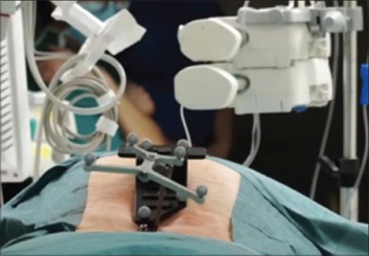
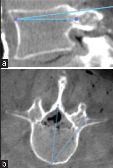
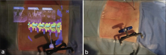
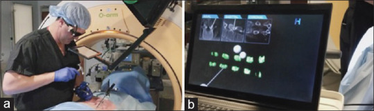
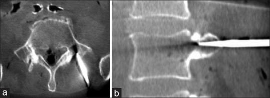
An augmented reality and artificial intelligence (ARAI)-assisted surgical navigation system was developed. The system consists of a display system which hovers over the surgical field and projects three-dimensional (3D) medical images corresponding with the patient's anatomy. The system was registered to the cadaveric spine using an optical tracker and arrays with reflective markers. The virtual image overlay from the ARAI system was compared to 3D generated images from intraoperative scans and used to percutaneously navigate a probe to the cortex at the corresponding pedicle starting point. Intraoperative scan was used to confirm the probe position. Virtual probe placement was compared to the actual probe position in the bone to determine the accuracy of the navigation system.
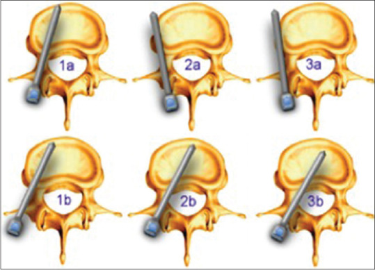
Four cadaveric thoracolumbar spines were used. The navigated probes were correctly placed in all attempted levels ( = 24 levels), defined as Zdichavsky type 1a, Ravi type I, and Gertzbein type 0. The virtual overlay image corresponded to the 3D generated image in all the tested levels.
The ARAI surgical navigation system correctly and accurately identified the starting points at all the attempted levels. The virtual anatomy image overlay precisely corresponded to the actual anatomy in all the tested scenarios. This technology may lead more uniform outcomes between surgeons and decrease minimally invasive spine surgery learning curves.
利用术中获取的计算机断层扫描生成的DICOM图像,基于增强现实的虚拟脊柱骨解剖图像叠加可投影到真实的脊柱解剖结构上。本研究的目的是开发一种技术,并评估使用增强现实辅助手术导航进行腰椎椎弓根器械置入的准确性和可行性。
开发了一种增强现实与人工智能(ARAI)辅助手术导航系统。该系统由一个显示系统组成,该显示系统悬浮在手术区域上方,并投射与患者解剖结构相对应的三维(3D)医学图像。使用光学跟踪器和带有反射标记的阵列将系统注册到尸体脊柱上。将ARAI系统的虚拟图像叠加与术中扫描生成的3D图像进行比较,并用于经皮将探针导航到相应椎弓根起始点的皮质。术中扫描用于确认探针位置。将虚拟探针放置与骨内实际探针位置进行比较,以确定导航系统的准确性。
使用了四个尸体胸腰椎脊柱。在所有尝试的节段(=24个节段)中,导航探针均正确放置,定义为Zdichavsky 1a型、Ravi I型和Gertzbein 0型。在所有测试节段中,虚拟叠加图像与3D生成图像相对应。
ARAI手术导航系统在所有尝试的节段中均正确且准确地识别了起始点。在所有测试场景中,虚拟解剖图像叠加与实际解剖结构精确对应。这项技术可能会使外科医生之间的手术效果更加一致,并降低微创脊柱手术的学习曲线。