Department of Chemical Engineering, University of Utah, Salt Lake City, Utah, USA.
Division of Otolaryngology-Head and Neck Surgery, University of Utah, Salt Lake City, Utah, USA.
Otolaryngol Head Neck Surg. 2021 Mar;164(3):547-555. doi: 10.1177/0194599820957966. Epub 2020 Sep 15.
To determine whether common otolaryngology procedures generate viable aerosolized virus through a murine cytomegalovirus (mCMV) model for infection.
mCMV model of infection.
University of Utah laboratory.
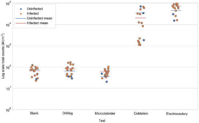
Three-day-old BALB/c mice were inoculated with mCMV or saline. Five days later, each mouse underwent drilling, microdebrider, coblation, and electrocautery procedures. Particle size distribution and PM (particulate matter <2.5 µm) concentration were determined with a scanning mobility particle sizer and an aerosol particle sizer in the range of 15 nm to 32 µm. Aerosolized samples from these procedures were collected with an Aerosol Devices BioSpot sampler for viral titer based on polymerase chain reaction and for viable virus through viral culture.
As compared with the background aerosol concentrations, coblation and electrocautery showed statistically significant increases in airborne aerosols (Tukey-adjusted value <.040), while microdebrider and drilling at 30,000 rpm did not (.870 < Tukey-adjusted value < .930). We identified viral DNA in samples from coblation and drilling procedures, although we did not identify viable viruses in aerosol samples from any of the 4 procedures.
Coblation and electrocautery procedures generate >100-fold increases in aerosol concentrations over background; only coblation and drilling produce aerosolized viral DNA. The high concentration of aerosols from coblation and electrocautery suggests the need for appropriate safeguards against particle exposure to health care workers. The presence of viral DNA from drilling and coblation procedures warrants the need for appropriate protection against droplet and aerosol exposure.
通过鼠巨细胞病毒 (mCMV) 感染模型,确定常见耳鼻喉科手术是否会产生具有感染力的气溶胶病毒。
mCMV 感染模型。
犹他大学实验室。
将 3 日龄 BALB/c 小鼠接种 mCMV 或生理盐水。5 天后,每只小鼠接受钻孔、微磨钻、等离子刀和电烙术。使用扫描迁移率颗粒分析仪和气溶胶颗粒分析仪,在 15nm 至 32µm 的范围内,测定粒径分布和 PM(颗粒物<2.5µm)浓度。对这些程序产生的气溶胶进行收集,使用 Aerosol Devices BioSpot 采样器进行基于聚合酶链反应的病毒滴度检测,以及通过病毒培养检测活病毒。
与背景气溶胶浓度相比,等离子刀和电烙术显示出统计学上显著增加的空气传播气溶胶(经 Tukey 调整后 P 值<.040),而微磨钻和 30000rpm 的钻孔则没有(.870<经 Tukey 调整后 P 值<.930)。我们在等离子刀和钻孔程序的样本中发现了病毒 DNA,但在任何 4 种程序的气溶胶样本中都未发现有活性的病毒。
等离子刀和电烙术程序产生的气溶胶浓度比背景高出 100 倍以上;只有等离子刀和钻孔程序产生了气溶胶化的病毒 DNA。等离子刀和电烙术产生的高浓度气溶胶提示需要采取适当的防护措施,防止医护人员接触颗粒。钻孔和等离子刀程序中存在病毒 DNA,这表明需要采取适当的防护措施,防止飞沫和气溶胶暴露。