Division in Anatomy and Developmental Biology, Department of Oral Biology, Human Identification Research Institute, BK21 PLUS Project, Yonsei University College of Dentistry, 50-1 Yonsei-ro, Seodaemun-gu, Seoul 03722, Korea.
Youth Clinic, 30 Apgujeong-ro 80-gil, Gangnam-gu, Seoul 03722, Korea.
Toxins (Basel). 2020 Sep 11;12(9):588. doi: 10.3390/toxins12090588.
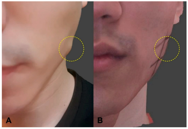
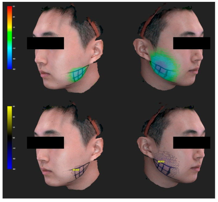
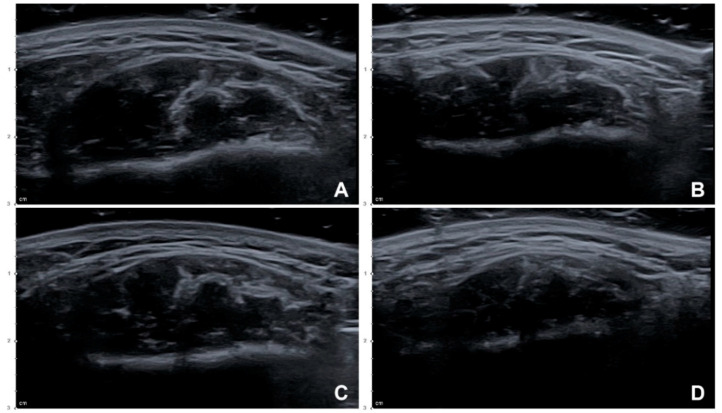
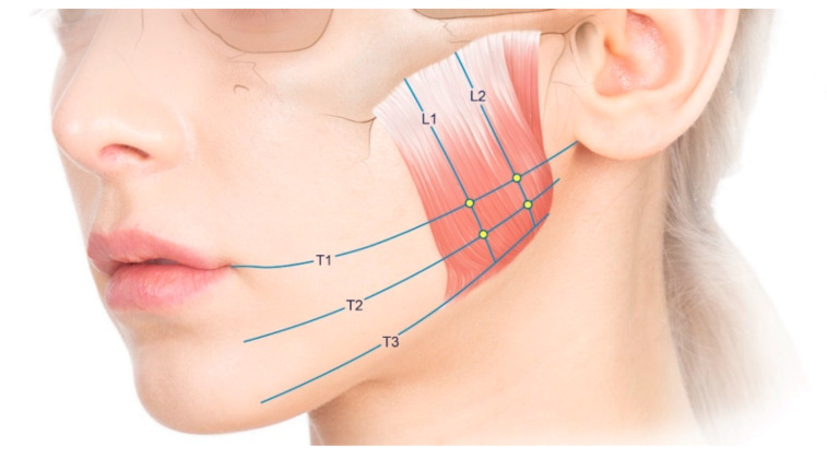
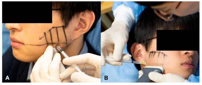
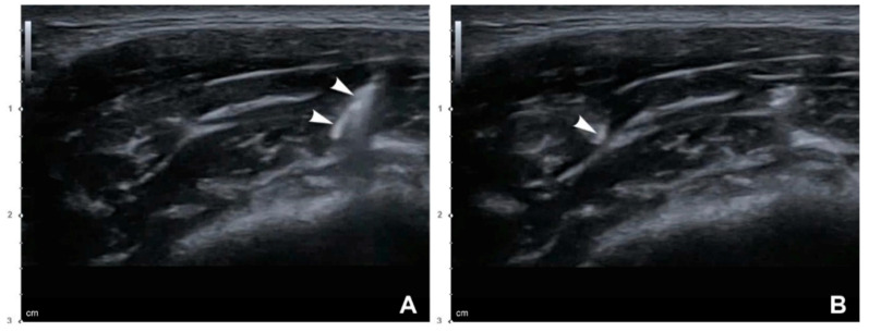
The aim of the study was to propose a more efficient and safer botulinum toxin type A (BoNT-A) injection method for the masseter by comparing the conventional blind injection and a novel ultrasonography (US)-guided injection technique in a clinical trial. The 40 masseters from 20 healthy young Korean volunteers (10 males and 10 females with a mean age of 25.6 years) were included in this prospective clinical trial. The BoNT-A (24 U) was injected into the masseter of each volunteer using the conventional blind and US-guided injection techniques on the left and right sides, respectively, and analyzed by US and three-dimensional (3D) facial scanning. One case of PMB (paradoxical masseteric bulging) was observed on the side where a conventional blind injection was performed, which disappeared after the compensational injection. The reduction in the thickness of the masseter in the resting state differed significantly at 1 month after the injection between the conventional blind injection group and the US-guided injection group by 12.38 ± 7.59% and 17.98 ± 9.65%, respectively ((19) = 3.059, = 0.007). The reduction in the facial contour also differed significantly at 1 month after the injection between the conventional blind injection group and the US-guided injection group by 1.95 ± 0.74 mm and 2.22 ± 0.84 mm, respectively ((19) = 2.908, = 0.009). The results of the study showed that the US-guided injection method that considers the deep inferior tendon by visualizing the masseter can prevent the PMB that can occur during a blind injection, and is also more effective.
本研究旨在通过临床试验比较传统盲注法和新型超声(US)引导注射法,提出一种更有效、更安全的咬肌肉毒毒素 A(BoNT-A)注射方法。该前瞻性临床试验纳入了 20 名健康年轻韩国志愿者(10 名男性和 10 名女性,平均年龄 25.6 岁)的 40 侧咬肌。志愿者两侧咬肌分别采用传统盲注法和 US 引导注射法,各注射 24U BoNT-A,采用 US 和三维(3D)面部扫描进行分析。1 例采用传统盲注法的志愿者出现了 PMB(反常性咬肌膨隆),补偿性注射后消失。注射后 1 个月,传统盲注组和 US 引导注射组在休息状态下咬肌厚度的减少分别为 12.38%±7.59%和 17.98%±9.65%,差异有统计学意义(t=-3.059,P=0.007)。注射后 1 个月,传统盲注组和 US 引导注射组在面部轮廓的减少分别为 1.95±0.74mm 和 2.22±0.84mm,差异有统计学意义(t=-2.908,P=0.009)。研究结果表明,通过可视化咬肌考虑深层下颌腱的 US 引导注射方法可以预防盲注时可能发生的 PMB,并且更有效。