Department of Anatomy and Structural Biology, Albert Einstein College of Medicine, Bronx, NY, USA.
Analytical Imaging Facility, Albert Einstein College of Medicine, Bronx, NY, USA.
Transl Vis Sci Technol. 2020 Aug 31;9(9):45. doi: 10.1167/tvst.9.9.45. eCollection 2020 Aug.
To use second harmonic generation imaging and fluorescence recovery after photobleaching to demonstrate alterations in scleral collagen structure and permeability after crosslinking in rat and human eyes.
Excised rat and human scleras were imaged ex vivo with an inverted two-photon excitation fluorescence microscope before and after photochemical crosslinking using riboflavin and 405-nm laser light. Fluorescence recovery after photobleaching was applied to measure the diffusion of fluorescein isothiocyanate-dextran across the sclera.
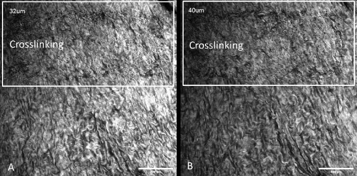
Crosslinking caused scleral collagen fibers to become wavier and more densely packed, with surface collagen being more affected than deeper collagen fibers. Crosslinked sclera showed significantly decreased permeability in the irradiation zone and also extended as far as 250 µm outside the irradiation zone.
Photochemical crosslinking induced changes in scleral structure and permeability that extended to tissue even outside the irradiation zone.
Ultrastructural changes associated with the emerging clinical technique of photochemical scleral crosslinking have not been well characterized. We demonstrate not only changes in scleral collagen by second harmonic generation imaging but also the associated functional changes in tissue permeability by fluorescence recovery after photobleaching. We report the novel finding of reduced permeability extending well beyond the direct irradiation zone. This has implications for control in the clinical setting.
利用二次谐波成像和光漂白后荧光恢复技术,证明在大鼠和人眼中交联后巩膜胶原结构和通透性的变化。
在使用核黄素和 405nm 激光进行光化学交联之前和之后,使用倒置双光子激发荧光显微镜对离体的大鼠和人巩膜进行成像。荧光漂白后荧光恢复用于测量异硫氰酸荧光素-葡聚糖穿过巩膜的扩散。
交联导致巩膜胶原纤维变得更加弯曲和密集,表面胶原比深层胶原纤维受影响更大。交联的巩膜在照射区域显示出明显降低的通透性,并且在照射区域外延伸了多达 250µm。
光化学交联引起的巩膜结构和通透性的变化延伸到组织中,甚至延伸到照射区域之外。
与光化学巩膜交联这一新兴临床技术相关的超微结构变化尚未得到很好的描述。我们不仅通过二次谐波成像显示了巩膜胶原的变化,还通过光漂白后荧光恢复显示了组织通透性的相关功能变化。我们报告了一个新的发现,即通透性降低的范围远远超出了直接照射区域。这对临床环境中的控制有影响。