• 文献检索
  • 文档翻译
  • 深度研究
  • 学术资讯
  • Suppr Zotero 插件Zotero 插件
  • 邀请有礼
  • 套餐&价格
  • 历史记录
应用&插件
Suppr Zotero 插件Zotero 插件浏览器插件Mac 客户端Windows 客户端微信小程序
定价
高级版会员购买积分包购买API积分包
服务
文献检索文档翻译深度研究API 文档MCP 服务
关于我们
关于 Suppr公司介绍联系我们用户协议隐私条款
关注我们

Suppr 超能文献

核心技术专利:CN118964589B侵权必究
粤ICP备2023148730 号-1Suppr @ 2026

文献检索

告别复杂PubMed语法,用中文像聊天一样搜索,搜遍4000万医学文献。AI智能推荐,让科研检索更轻松。

立即免费搜索

文件翻译

保留排版,准确专业,支持PDF/Word/PPT等文件格式,支持 12+语言互译。

免费翻译文档

深度研究

AI帮你快速写综述,25分钟生成高质量综述,智能提取关键信息,辅助科研写作。

立即免费体验

乳腺良恶性病变在对比增强数字化乳腺 X 线摄影中的动力学表现。

Kinetic patterns of benign and malignant breast lesions on contrast enhanced digital mammogram.

机构信息

Department of Radiology, Kaohsiung Veterans General Hospital, Kaohsiung, Taiwan, ROC.

National Yang-Ming University, School of Medicine, Taipei, Taiwan, ROC.

出版信息

PLoS One. 2020 Sep 17;15(9):e0239271. doi: 10.1371/journal.pone.0239271. eCollection 2020.

DOI:10.1371/journal.pone.0239271
PMID:32941537
原文链接:https://pmc.ncbi.nlm.nih.gov/articles/PMC7498093/
Abstract

PURPOSE

To evaluate the kinetic patterns of benign and malignant breast lesions using contrast-enhanced digital mammogram (CEDM).

METHODS

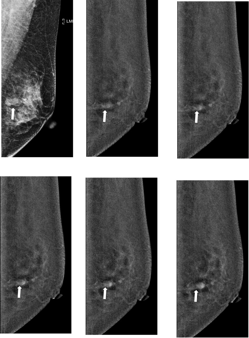
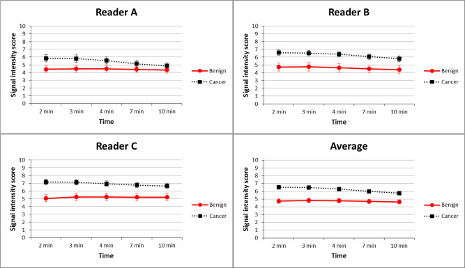
Women with suspicious breast lesions on mammography or ultrasound were enrolled. Single-view mediolateral oblique (MLO) CEDM of an affected breast was acquired at 2, 3, 4, 7, and 10 min after injection of contrast agent. Three readers visually and semi-quantitatively analyzed the enhancement of suspicious lesions. The kinetic pattern of each lesion was classified as persistent, plateau, or washout over two time intervals, 2-4 min and 2-10 min, by comparing the signal intensity at the first time interval with that at the second.

RESULTS

There were 73 malignant and 75 benign lesions in 148 patients (mean age: 52 years). Benign and malignant breast lesions showed the highest signal intensity at 3 min and 2 min, respectively. Average areas under receiver operating characteristic (ROC) curve for diagnostic accuracy based on lesion enhancement at different time points were 0.73 at 2 min, 0.72 at 3 min, 0.69 at 4 min, 0.67 at 7 min, and 0.64 at 10 min. Diagnostic performance was significantly better at 2, 3, and 4 min than at 7 and 10 min (all p < 0.05). A washout kinetic pattern was significantly associated with malignant lesions at 2-4 min and 2-10 min frames according to two of the three readers' interpretations (all p ≤ 0.001).

CONCLUSION

Applications of optimal time intervals and kinetic patterns show promise in differentiation of benign and malignant breast lesions on CEDM.

摘要

目的

利用对比增强数字乳腺断层摄影术(CEDM)评估良性和恶性乳腺病变的动力学模式。

方法

招募乳腺钼靶或超声检查发现可疑乳腺病变的女性。在注射造影剂后 2、3、4、7 和 10 分钟,对受影响的乳房进行单视图内外斜位(MLO)CEDM。三位读者分别进行视觉和半定量分析可疑病变的增强情况。通过比较第一个时间间隔和第二个时间间隔的信号强度,将每个病变的动力学模式分为持续、平台或廓清。

结果

148 例患者中有 73 例恶性和 75 例良性病变(平均年龄:52 岁)。良性和恶性乳腺病变在 3 分钟和 2 分钟时分别显示出最高的信号强度。基于不同时间点病变增强的诊断准确性的受试者工作特征(ROC)曲线下平均面积分别为 2 分钟时 0.73、3 分钟时 0.72、4 分钟时 0.69、7 分钟时 0.67、10 分钟时 0.64。在 2、3 和 4 分钟时,诊断性能明显优于 7 和 10 分钟(均 p<0.05)。根据三位读者中的两位的解释,在 2-4 分钟和 2-10 分钟时,廓清动力学模式与恶性病变显著相关(均 p≤0.001)。

结论

CEDM 上应用最佳时间间隔和动力学模式有望区分良性和恶性乳腺病变。

https://cdn.ncbi.nlm.nih.gov/pmc/blobs/3ad7/7498093/6e21111eca4e/pone.0239271.g006.jpg
https://cdn.ncbi.nlm.nih.gov/pmc/blobs/3ad7/7498093/b7f3320fc806/pone.0239271.g001.jpg
https://cdn.ncbi.nlm.nih.gov/pmc/blobs/3ad7/7498093/2d8df7faa3fe/pone.0239271.g002.jpg
https://cdn.ncbi.nlm.nih.gov/pmc/blobs/3ad7/7498093/27cf58f7ef64/pone.0239271.g003.jpg
https://cdn.ncbi.nlm.nih.gov/pmc/blobs/3ad7/7498093/555181be99a8/pone.0239271.g004.jpg
https://cdn.ncbi.nlm.nih.gov/pmc/blobs/3ad7/7498093/282fc50ea9d8/pone.0239271.g005.jpg
https://cdn.ncbi.nlm.nih.gov/pmc/blobs/3ad7/7498093/6e21111eca4e/pone.0239271.g006.jpg

相似文献

1
Kinetic patterns of benign and malignant breast lesions on contrast enhanced digital mammogram.乳腺良恶性病变在对比增强数字化乳腺 X 线摄影中的动力学表现。
PLoS One. 2020 Sep 17;15(9):e0239271. doi: 10.1371/journal.pone.0239271. eCollection 2020.
2
Clinical evaluation of contrast-enhanced digital mammography and contrast enhanced tomosynthesis--Comparison to contrast-enhanced breast MRI.对比增强数字化乳腺摄影和对比增强断层合成的临床评估——与对比增强乳腺MRI的比较。
Eur J Radiol. 2015 Dec;84(12):2501-8. doi: 10.1016/j.ejrad.2015.09.019. Epub 2015 Oct 1.
3
Dual-energy contrast-enhanced digital mammography: initial clinical results of a multireader, multicase study.双能数字乳腺造影术:一项多读者、多病例研究的初步临床结果。
Breast Cancer Res. 2012 Jun 14;14(3):R94. doi: 10.1186/bcr3210.
4
Differentiation between benign and malignant breast tumors using kinetic features of real-time harmonic contrast-enhanced ultrasound.利用实时谐波对比增强超声的动力学特征鉴别乳腺良恶性肿瘤
Acta Radiol. 2012 May 1;53(4):382-8. doi: 10.1258/ar.2012.110562. Epub 2012 Mar 20.
5
Correlation between quantitative assessment of contrast enhancement in contrast-enhanced spectral mammography (CESM) and histopathology-preliminary results.对比增强光谱乳腺摄影术(CESM)中对比增强定量评估与组织病理学的相关性——初步结果。
Eur Radiol. 2019 Nov;29(11):6220-6226. doi: 10.1007/s00330-019-06232-6. Epub 2019 May 14.
6
Quantitative analysis of enhanced malignant and benign lesions on contrast-enhanced spectral mammography.对比增强光谱乳腺摄影术中良恶性病变强化的定量分析。
Br J Radiol. 2018 Jun;91(1086):20170605. doi: 10.1259/bjr.20170605. Epub 2018 Feb 27.
7
Dual-energy contrast-enhanced digital mammography: initial clinical results.双能数字化乳腺造影:初步临床结果。
Eur Radiol. 2011 Mar;21(3):565-74. doi: 10.1007/s00330-010-1944-y. Epub 2010 Sep 14.
8
Improving suspicious breast lesion characterization using semi-automatic lesion fractional volume washout kinetic analysis.利用半自动病变分容积洗脱动力学分析提高可疑乳腺病变的特征描述。
Med Phys. 2011 Nov;38(11):5998-6009. doi: 10.1118/1.3651635.
9
Diagnostic value of qualitative and quantitative enhancement parameters on contrast-enhanced mammography.定性和定量增强参数在对比增强乳腺摄影中的诊断价值。
Diagn Interv Radiol. 2024 Jul 8;30(4):248-255. doi: 10.4274/dir.2024.232472. Epub 2024 Apr 15.
10
Preoperative loco-regional staging of invasive lobular carcinoma with contrast-enhanced digital mammography (CEDM).术前对比增强数字乳腺摄影术(CEDM)对浸润性小叶癌的局部区域分期。
Radiol Med. 2019 Dec;124(12):1229-1237. doi: 10.1007/s11547-019-01116-7. Epub 2019 Nov 26.

引用本文的文献

1
Efficacy of Contrast-Enhanced Mammography in the Evaluation of Breast Lesions: An Initial Experience.对比增强乳腺钼靶摄影在乳腺病变评估中的效能:初步经验
Cureus. 2025 Jul 8;17(7):e87566. doi: 10.7759/cureus.87566. eCollection 2025 Jul.
2
Addition of contrast-enhanced mammography enhancement patterns and morphology for differentiating benign from malignant papillary breast lesions.增加对比增强乳腺X线摄影的强化模式和形态学特征以鉴别乳腺乳头状病变的良恶性。
Br J Radiol. 2025 Mar 1;98(1167):383-391. doi: 10.1093/bjr/tqae241.
3
Contrast-enhanced mammography improves patient access to functional breast imaging.

本文引用的文献

1
Performance of Dual-Energy Contrast-enhanced Digital Mammography for Screening Women at Increased Risk of Breast Cancer.双能增强数字乳腺摄影筛查乳腺癌高危女性的性能。
Radiology. 2019 Oct;293(1):81-88. doi: 10.1148/radiol.2019182660. Epub 2019 Aug 27.
2
Evaluation of single-view contrast-enhanced mammography as novel reading strategy: a non-inferiority feasibility study.单视图对比增强乳腺摄影作为新型阅读策略的评估:一项非劣效性可行性研究。
Eur Radiol. 2019 Nov;29(11):6211-6219. doi: 10.1007/s00330-019-06215-7. Epub 2019 May 9.
3
Comparison of Upright Digital Breast Tomosynthesis-guided versus Prone Stereotactic Vacuum-assisted Breast Biopsy.
对比增强乳腺钼靶摄影改善了患者获得功能性乳腺成像的机会。
J Med Imaging Radiat Oncol. 2025 Feb;69(1):46-61. doi: 10.1111/1754-9485.13789. Epub 2024 Oct 31.
4
Enhancing the Understanding of Breast Vascularity Through Insights From Dynamic Contrast-Enhanced Magnetic Resonance Imaging: A Comprehensive Review.通过动态对比增强磁共振成像的见解加深对乳腺血管的理解:一项全面综述
Cureus. 2024 Sep 26;16(9):e70226. doi: 10.7759/cureus.70226. eCollection 2024 Sep.
5
Application of the Kaiser score on contrast-enhanced mammography in the differential diagnosis of breast lesions: comparison with breast magnetic resonance imaging.凯泽评分在乳腺造影增强检查中对乳腺病变鉴别诊断的应用:与乳腺磁共振成像的比较
Quant Imaging Med Surg. 2024 Aug 1;14(8):5541-5554. doi: 10.21037/qims-24-593. Epub 2024 Jul 18.
6
Role of contrast-enhanced mammogram as an adjunct to tomosynthesis in evaluation of circumscribed breast lesions.对比增强乳腺 X 线摄影在评估局限性乳腺病变中的作用。
Br J Radiol. 2024 Oct 1;97(1162):1696-1705. doi: 10.1093/bjr/tqae130.
7
Nomogram for preoperative differentiation of benign and malignant breast tumors using contrast-enhanced cone-beam breast CT (CE CB-BCT) quantitative imaging and assessment features.基于对比增强锥形束乳腺 CT(CE CB-BCT)定量成像和评估特征的术前鉴别乳腺良恶性肿瘤的列线图。
Radiol Med. 2024 May;129(5):737-750. doi: 10.1007/s11547-024-01803-0. Epub 2024 Mar 21.
8
Contrast-enhanced spectral mammography: are kinetic patterns useful for differential diagnoses of enhanced lesions?对比增强光谱乳腺摄影:动力学模式对增强病变的鉴别诊断是否有用?
Diagn Interv Radiol. 2023 Mar 29;29(2):244-250. doi: 10.5152/dir.2022.21562. Epub 2023 Feb 27.
9
Can the delayed phase of quantitative contrast-enhanced mammography improve the diagnostic performance on breast masses?乳腺X线定量对比增强检查的延迟期能否提高乳腺肿块的诊断效能?
Quant Imaging Med Surg. 2021 Aug;11(8):3684-3697. doi: 10.21037/qims-20-1092.
直立式数字化乳腺断层摄影引导与俯卧式立体定位真空辅助乳腺活检的比较。
Radiology. 2019 Feb;290(2):298-304. doi: 10.1148/radiol.2018181788. Epub 2018 Dec 4.
4
Comparison of screening CEDM and MRI for women at increased risk for breast cancer: A pilot study.比较 CEDM 筛查与 MRI 筛查对乳腺癌高危女性的效果:一项初步研究。
Eur J Radiol. 2017 Dec;97:37-43. doi: 10.1016/j.ejrad.2017.10.001. Epub 2017 Oct 7.
5
Breast cancer screening with digital breast tomosynthesis - 4 year experience and comparison with national data.数字乳腺断层合成筛查乳腺癌-4 年的经验及与全国数据的比较。
J Chin Med Assoc. 2018 Jan;81(1):70-80. doi: 10.1016/j.jcma.2017.05.013. Epub 2017 Nov 9.
6
Susceptibility of iodine concentration map of dual-energy contrast-enhanced digital mammography for quantitative and tumor enhancement assessment.双能对比增强数字乳腺摄影碘浓度图在定量及肿瘤强化评估中的敏感性
Acta Radiol. 2018 Aug;59(8):893-901. doi: 10.1177/0284185117740760. Epub 2017 Nov 8.
7
Clinical evaluation of contrast-enhanced digital mammography and contrast enhanced tomosynthesis--Comparison to contrast-enhanced breast MRI.对比增强数字化乳腺摄影和对比增强断层合成的临床评估——与对比增强乳腺MRI的比较。
Eur J Radiol. 2015 Dec;84(12):2501-8. doi: 10.1016/j.ejrad.2015.09.019. Epub 2015 Oct 1.
8
Alternative screening for dense breasts: MRI.致密乳腺的替代筛查方法:磁共振成像(MRI)。
AJR Am J Roentgenol. 2015 Feb;204(2):W141-9. doi: 10.2214/AJR.14.13636.
9
Dual-energy contrast-enhanced digital mammography: initial clinical results.双能数字化乳腺造影:初步临床结果。
Eur Radiol. 2011 Mar;21(3):565-74. doi: 10.1007/s00330-010-1944-y. Epub 2010 Sep 14.
10
Pure ductal carcinoma in situ: a range of MRI features.单纯导管原位癌:一系列磁共振成像特征
AJR Am J Roentgenol. 2008 Sep;191(3):689-99. doi: 10.2214/AJR.07.3779.