Department of Radiology, Institut de cancérologie Gustave-Roussy, 39 rue Camille Desmoulin, Villejuif, 94805 France.
Breast Cancer Res. 2012 Jun 14;14(3):R94. doi: 10.1186/bcr3210.
The purpose of this study was to compare the diagnostic accuracy of dual-energy contrast-enhanced digital mammography (CEDM) as an adjunct to mammography (MX) ± ultrasonography (US) with the diagnostic accuracy of MX ± US alone.
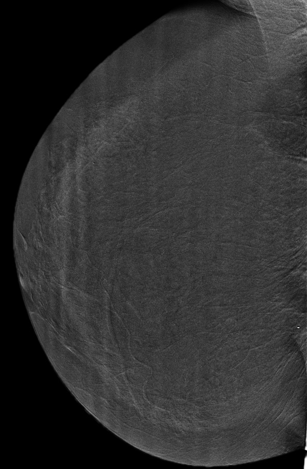
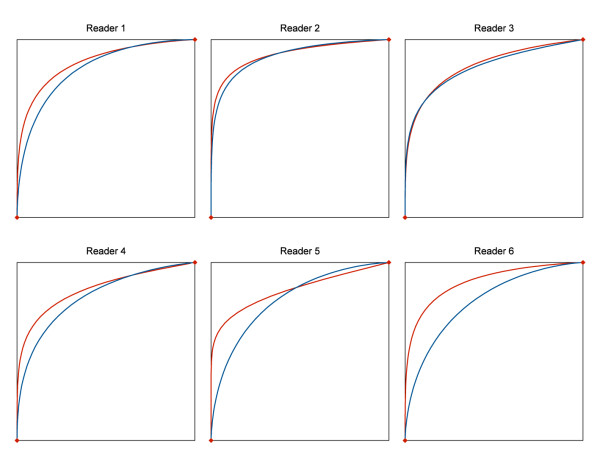
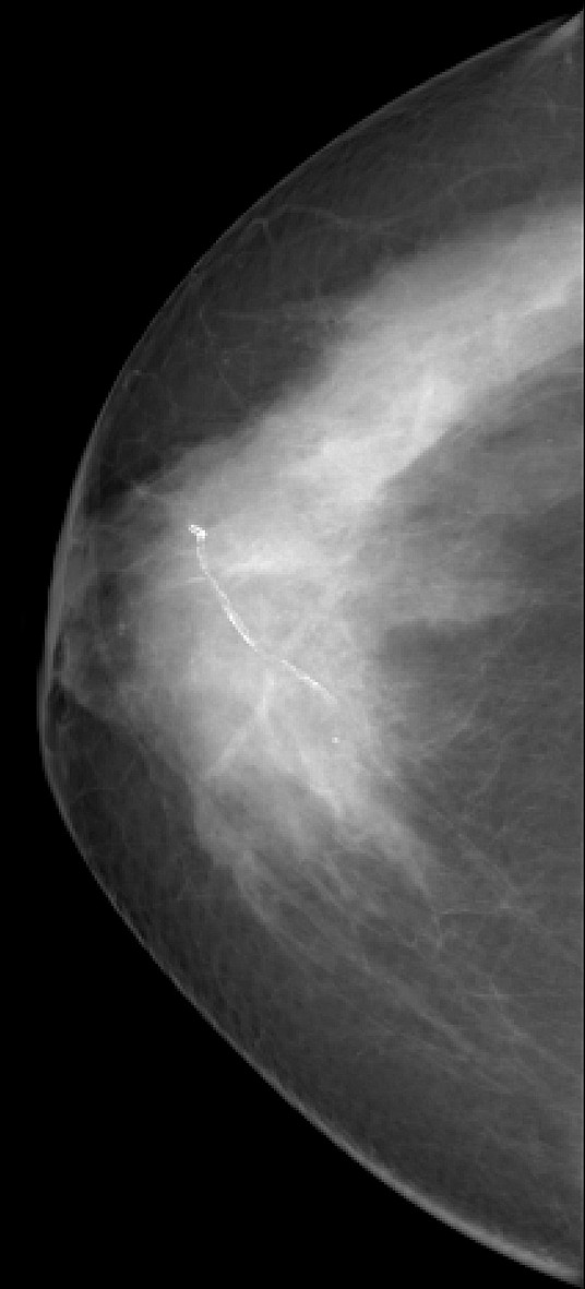
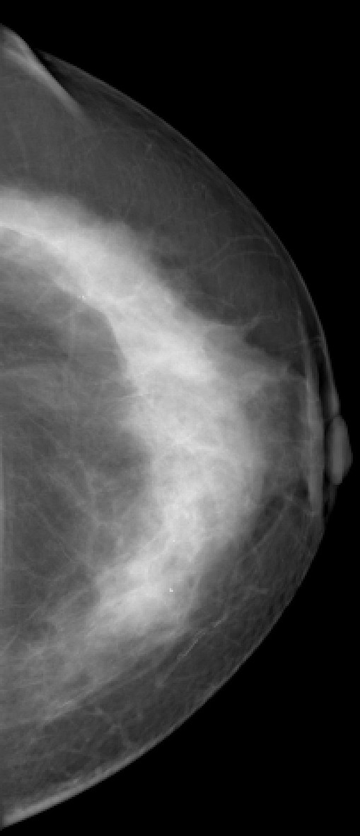
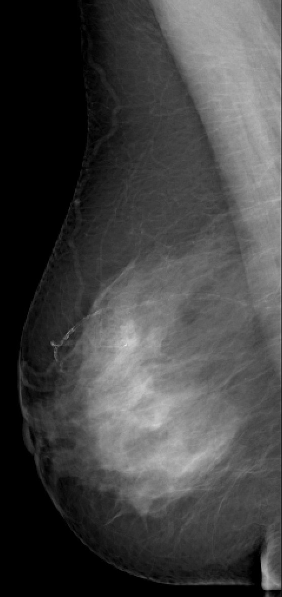
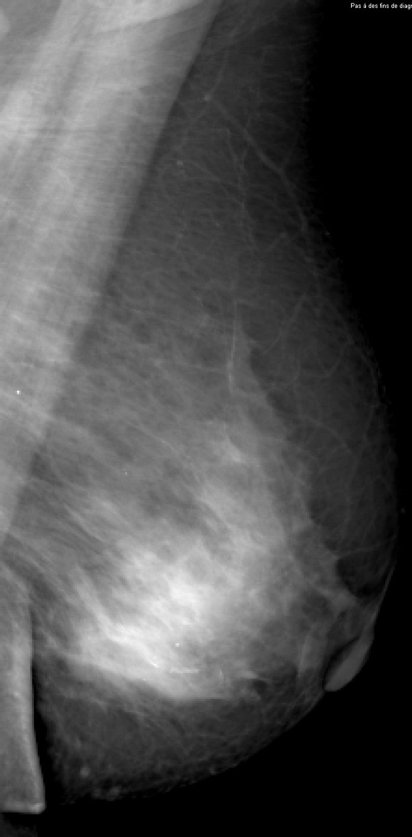
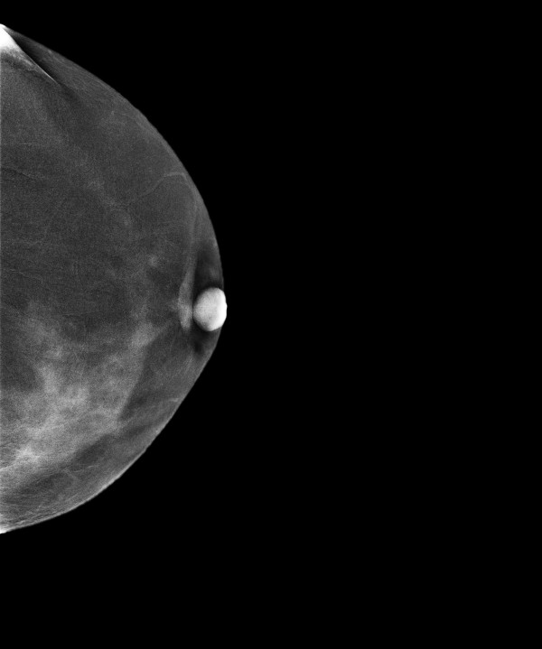
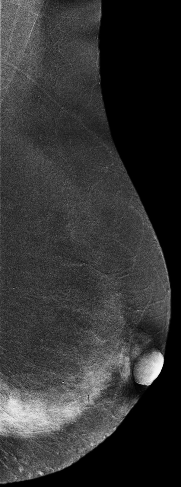
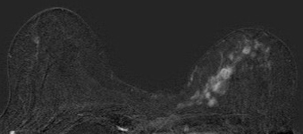
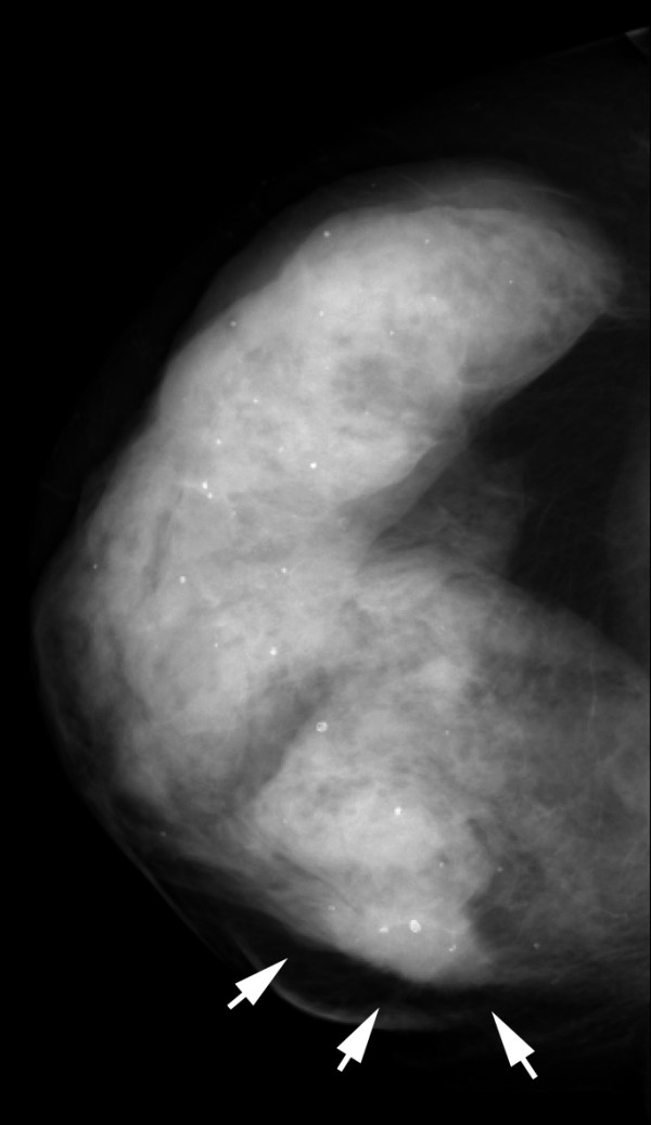
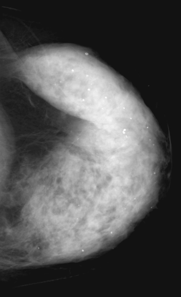
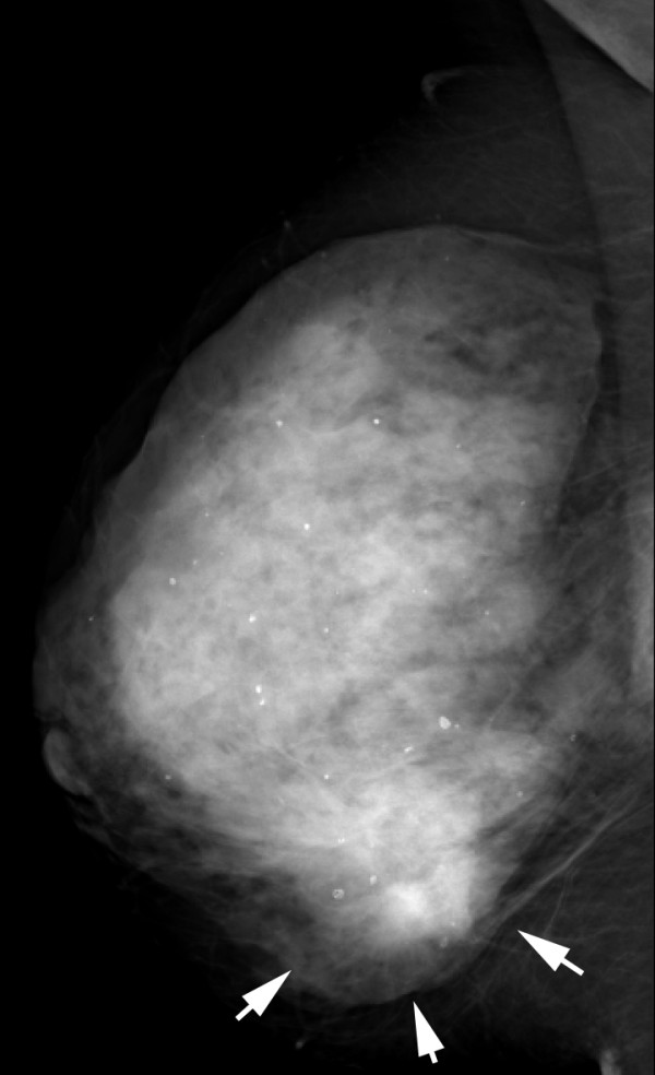
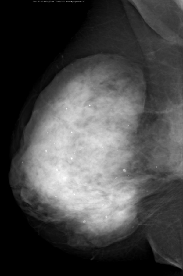
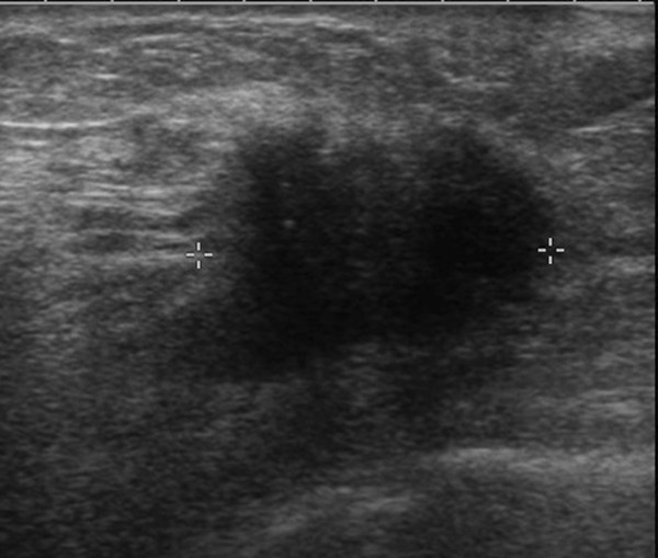
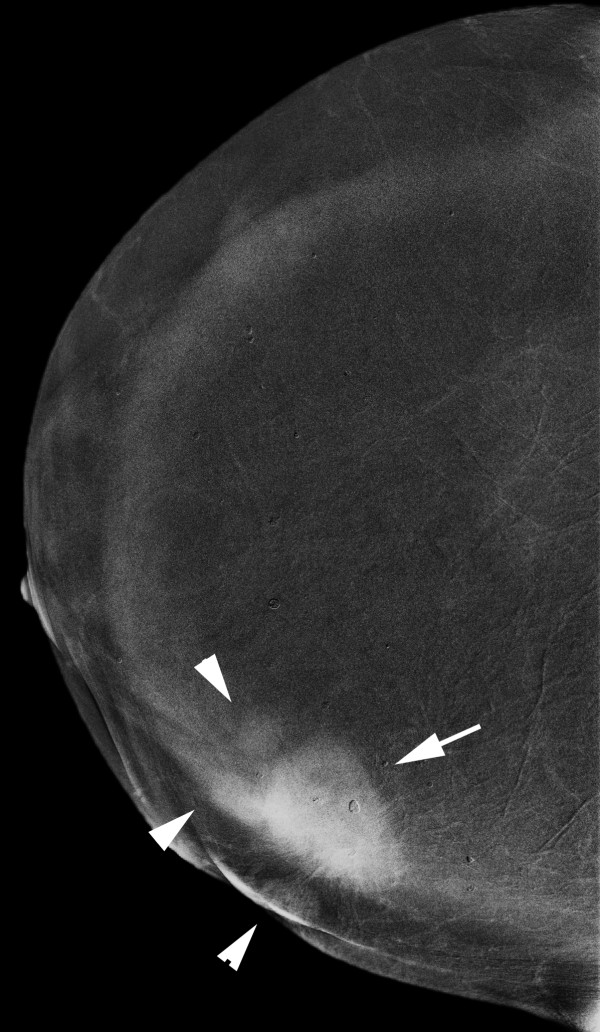
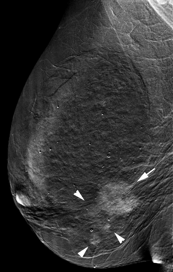
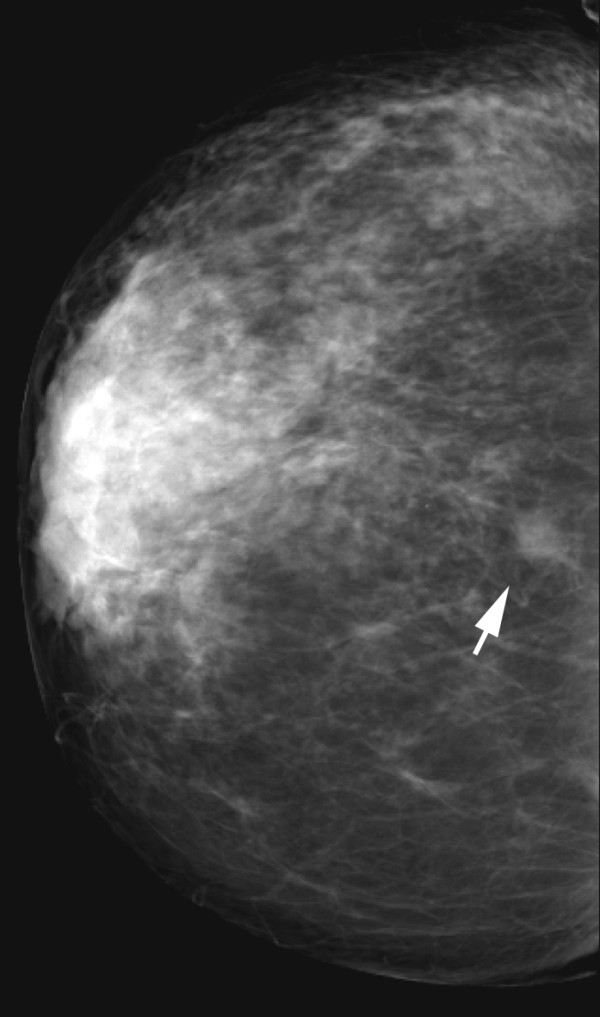
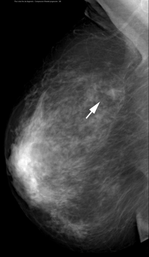
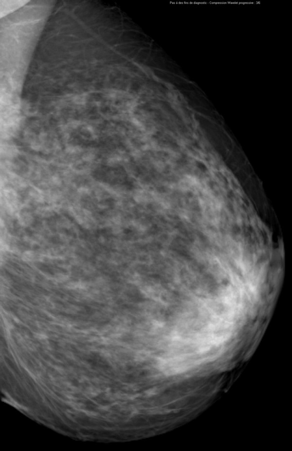
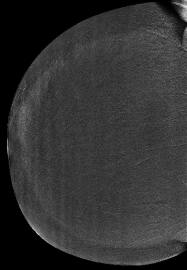
One hundred ten consenting women with 148 breast lesions (84 malignant, 64 benign) underwent two-view dual-energy CEDM in addition to MX and US using a specially modified digital mammography system (Senographe DS, GE Healthcare). Reference standard was histology for 138 lesions and follow-up for 12 lesions. Six radiologists from 4 institutions interpreted the images using high-resolution softcopy workstations. Confidence of presence (5-point scale), probability of cancer (7-point scale), and BI-RADS scores were evaluated for each finding. Sensitivity, specificity and ROC curve areas were estimated for each reader and overall. Visibility of findings on MX ± CEDM and MX ± US was evaluated with a Likert scale.
The average per-lesion sensitivity across all readers was significantly higher for MX ± US ± CEDM than for MX ± US (0.78 vs. 0.71 using BIRADS, p = 0.006). All readers improved their clinical performance and the average area under the ROC curve was significantly superior for MX ± US ± CEDM than for MX ± US ((0.87 vs 0.83, p = 0.045). Finding visibility was similar or better on MX ± CEDM than MX ± US in 80% of cases.
Dual-energy contrast-enhanced digital mammography as an adjunct to MX ± US improves diagnostic accuracy compared to MX ± US alone. Addition of iodinated contrast agent to MX facilitates the visualization of breast lesions.
本研究旨在比较双能对比增强数字乳腺摄影(CEDM)作为乳腺摄影(MX)±超声(US)辅助检查与单独 MX±US 的诊断准确性。
110 名同意参加研究的女性共 148 个乳腺病变(84 个恶性,64 个良性)接受了双能 CEDM 两视图检查,同时还进行了 MX 和 US 检查,使用了特殊改装的数字乳腺摄影系统(Senographe DS,GE Healthcare)。138 个病变的参考标准为组织学,12 个病变的参考标准为随访。来自 4 个机构的 6 名放射科医生使用高分辨率软拷贝工作站对图像进行了评估。对每个发现的存在信心(5 分制)、癌症可能性(7 分制)和 BI-RADS 评分进行了评估。估计了每位读者和总体的敏感性、特异性和 ROC 曲线面积。使用李克特量表评估了 MX±CEDM 和 MX±US 上发现的可见性。
所有读者的平均每病变敏感性,MX±US±CEDM 均明显高于 MX±US(使用 BI-RADS,p=0.006,0.78 对 0.71)。所有读者都提高了他们的临床性能,MX±US±CEDM 的平均 ROC 曲线下面积明显优于 MX±US(0.87 对 0.83,p=0.045)。在 80%的病例中,MX±CEDM 上的发现可见性与 MX±US 相似或更好。
与单独使用 MX±US 相比,作为 MX±US 辅助检查的双能对比增强数字乳腺摄影可提高诊断准确性。在 MX 中加入碘造影剂有助于可视化乳腺病变。