Department of Laboratory Medicine, The First Affiliated Hospital of Wenzhou Medical University, Wenzhou, Zhejiang 325027, P.R. China.
Department of Intensive Care Unit, The First Affiliated Hospital of Wenzhou Medical University, Wenzhou, Zhejiang 325027, P.R. China.
Oncol Rep. 2020 Oct;44(4):1616-1626. doi: 10.3892/or.2020.7721. Epub 2020 Aug 7.
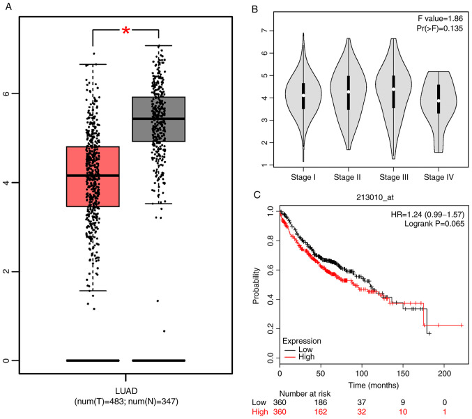
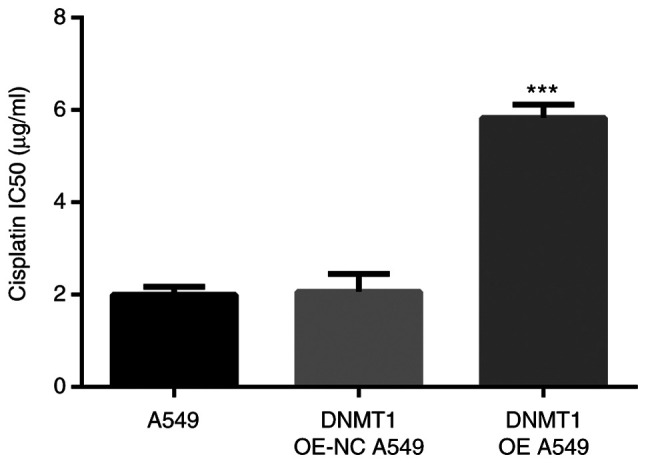
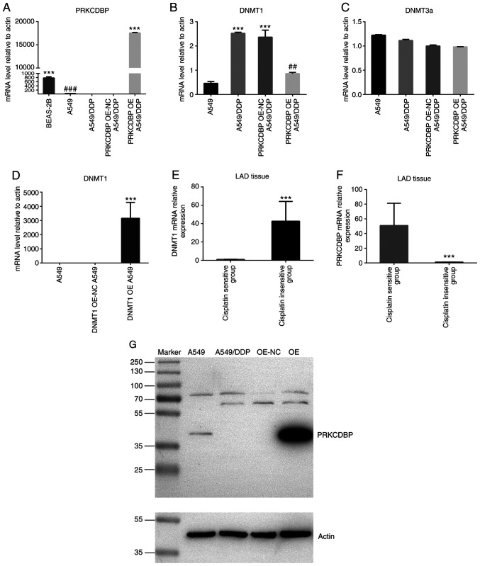
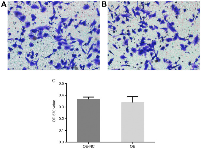
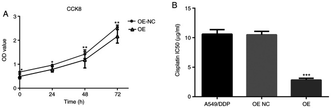
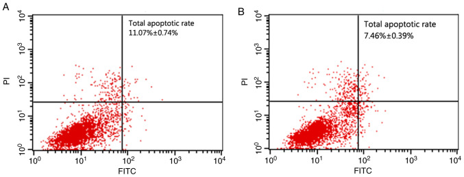
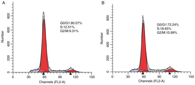
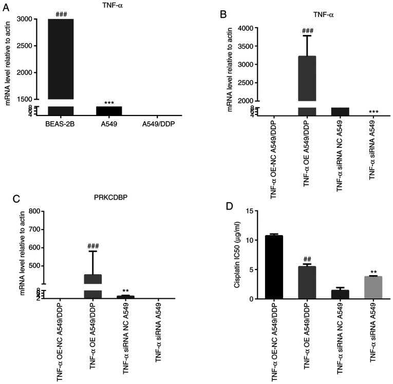
The aim of the present study was to explore the mechanism of protein kinase C delta binding protein (PRKCDBP) promoting cisplatin resistance in lung adenocarcinoma (LAD). The PRKCDBP expression level was herein detected by reverse transcription‑quantitative polymerase chain reaction (RT‑qPCR). We overexpressed PRKCDBP and tumor necrosis factor‑α (TNF‑α) in A549/DDP cell line, DNMT1 in A549 cells and siRNA TNF‑α in A549 cells with lentivirus‑mediated technique, and then, analyzed their biological diversification. The results showed a significantly lower expression level of PRKCDBP was lowly expressed in the A549/DDP cell line and LAD tissues than that in A549 cells and adjacent cancer tissues (P<0.05 and P<0.01), while the DNMT1 mRNA level was remarkably increased (P=0.000) and the promoter of PRKCDBP was hypermethylated in the A549/DDP cell line. Additionally, DNMT1 mRNA level in cisplatin‑insensitive group was markedly higher than that in cisplatin‑sensitive group (t=7.233, P<0.0001), while PRKCDBP mRNA level in cisplatin insensitive group was notably lower than that in cisplatin‑sensitive group (t=8.784, P<0.0001). The results showed that PRKCDBP mRNA level was significantly elevated following treatment with 5 µM decitabine for 24 h (P<0.0001), while the DNMT1 mRNA level was notably reduced (P=0.000). When PRKCDBP was overexpressed, the DNMT1 mRNA level was markedly decreased (P=0.007), the rate of proliferation (P<0.05 or P<0.01), IC50 of cisplatin (P<0.001), G2/M phase and S phase cells were obviously reduced (P<0.001), while G0/G1 phase cells, apoptosis (P<0.001) distinctly increased, but migration ability did not significantly change. TNF‑α overexpression resulted in an increase of PRKCDBP mRNA level (P<0.001), while TNF‑α siRNA led to PRKCDBP mRNA level distinctly reduced (P<0.001). Overexpression of DNMT1 improved IC50 in A549 cells. Thus, findings of the present study ascertained the promoter of PRKCDBP was hypermethylated in A549/DDP cells. In conclusion, low expression of PRKCDBP promoted cisplatin resistance in LAD by DNMT1 and TNF‑α.
本研究旨在探讨蛋白激酶 C 德尔塔结合蛋白(PRKCDBP)促进肺腺癌(LAD)顺铂耐药的机制。通过逆转录-定量聚合酶链反应(RT-qPCR)检测 PRKCDBP 的表达水平。我们通过慢病毒介导的技术过表达 A549/DDP 细胞系中的 PRKCDBP 和肿瘤坏死因子-α(TNF-α)、A549 细胞中的 DNMT1 和 A549 细胞中的 siRNA TNF-α,然后分析它们的生物学多样性。结果显示,A549/DDP 细胞系和 LAD 组织中 PRKCDBP 的表达水平明显低于 A549 细胞和相邻癌组织(P<0.05 和 P<0.01),而 DNMT1 mRNA 水平显著升高(P=0.000),PRKCDBP 启动子在 A549/DDP 细胞系中发生超甲基化。此外,顺铂耐药组的 DNMT1 mRNA 水平明显高于顺铂敏感组(t=7.233,P<0.0001),而顺铂耐药组的 PRKCDBP mRNA 水平明显低于顺铂敏感组(t=8.784,P<0.0001)。结果表明,用 5μM 地西他滨处理 24 小时后,PRKCDBP mRNA 水平显著升高(P<0.0001),而 DNMT1 mRNA 水平明显降低(P=0.000)。过表达 PRKCDBP 后,DNMT1 mRNA 水平明显降低(P=0.007),增殖率(P<0.05 或 P<0.01)、顺铂的 IC50(P<0.001)、G2/M 期和 S 期细胞明显减少(P<0.001),而 G0/G1 期细胞、凋亡(P<0.001)明显增加,而迁移能力没有明显变化。TNF-α 过表达导致 PRKCDBP mRNA 水平升高(P<0.001),而 TNF-α siRNA 导致 PRKCDBP mRNA 水平明显降低(P<0.001)。DNMT1 的过表达提高了 A549 细胞的 IC50。因此,本研究的结果证实 A549/DDP 细胞中 PRKCDBP 的启动子发生了超甲基化。总之,低表达 PRKCDBP 通过 DNMT1 和 TNF-α 促进 LAD 对顺铂的耐药性。