• 文献检索
  • 文档翻译
  • 深度研究
  • 学术资讯
  • Suppr Zotero 插件Zotero 插件
  • 邀请有礼
  • 套餐&价格
  • 历史记录
应用&插件
Suppr Zotero 插件Zotero 插件浏览器插件Mac 客户端Windows 客户端微信小程序
定价
高级版会员购买积分包购买API积分包
服务
文献检索文档翻译深度研究API 文档MCP 服务
关于我们
关于 Suppr公司介绍联系我们用户协议隐私条款
关注我们

Suppr 超能文献

核心技术专利:CN118964589B侵权必究
粤ICP备2023148730 号-1Suppr @ 2026

文献检索

告别复杂PubMed语法,用中文像聊天一样搜索,搜遍4000万医学文献。AI智能推荐,让科研检索更轻松。

立即免费搜索

文件翻译

保留排版,准确专业,支持PDF/Word/PPT等文件格式,支持 12+语言互译。

免费翻译文档

深度研究

AI帮你快速写综述,25分钟生成高质量综述,智能提取关键信息,辅助科研写作。

立即免费体验

在小鼠中,通过肾上主动脉缩窄造成的压力超负荷会导致左心室肥厚,而心肌细胞中没有 c-Kit 的表达。

Pressure overload by suprarenal aortic constriction in mice leads to left ventricular hypertrophy without c-Kit expression in cardiomyocytes.

机构信息

Division of Molecular Cardiology and Biophysics, Victor Chang Cardiac Research Institute, 405 Liverpool Street, Darlinghurst, Sydney, NSW, 2010, Australia.

St Vincent's Clinical School, University of New South Wales, Sydney, NSW, 2052, Australia.

出版信息

Sci Rep. 2020 Sep 18;10(1):15318. doi: 10.1038/s41598-020-72273-3.

DOI:10.1038/s41598-020-72273-3
PMID:32948799
原文链接:https://pmc.ncbi.nlm.nih.gov/articles/PMC7501855/
Abstract

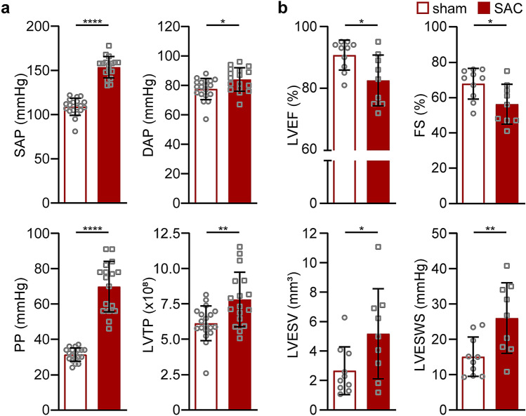
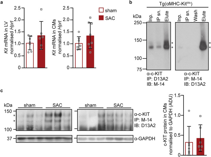
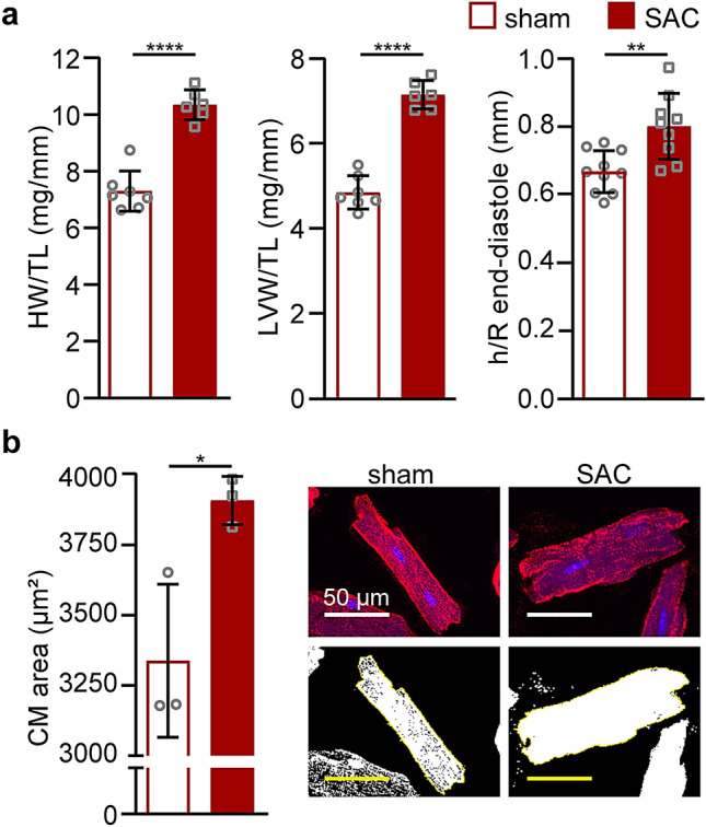
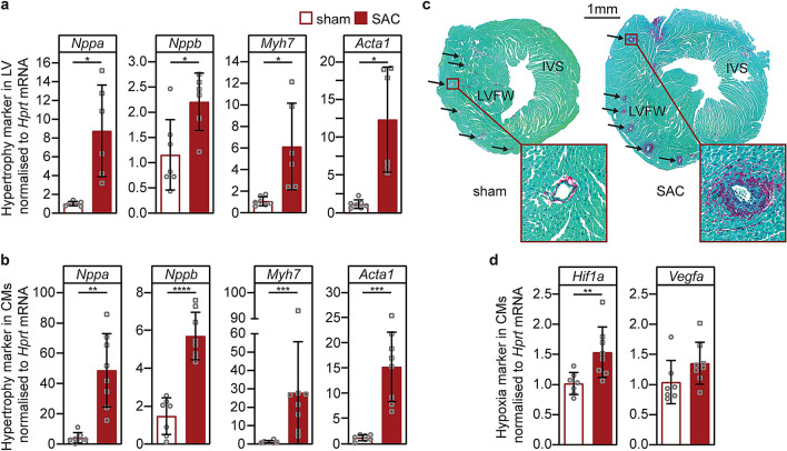
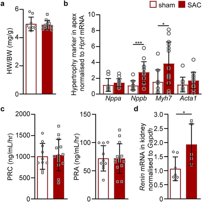
Animal models of pressure overload are valuable for understanding hypertensive heart disease. We characterised a surgical model of pressure overload-induced hypertrophy in C57BL/6J mice produced by suprarenal aortic constriction (SAC). Compared to sham controls, at one week post-SAC systolic blood pressure was significantly elevated and left ventricular (LV) hypertrophy was evident by a 50% increase in the LV weight-to-tibia length ratio due to cardiomyocyte hypertrophy. As a result, LV end-diastolic wall thickness-to-chamber radius (h/R) ratio increased, consistent with the development of concentric hypertrophy. LV wall thickening was not sufficient to normalise LV wall stress, which also increased, resulting in LV systolic dysfunction with reductions in ejection fraction and fractional shortening, but no evidence of heart failure. Pathological LV remodelling was evident by the re-expression of fetal genes and coronary artery perivascular fibrosis, with ischaemia indicated by enhanced cardiomyocyte Hif1a expression. The expression of stem cell factor receptor, c-Kit, was low basally in cardiomyocytes and did not change following the development of robust hypertrophy, suggesting there is no role for cardiomyocyte c-Kit signalling in pathological LV remodelling following pressure overload.

摘要

动物压力超负荷模型对于理解高血压性心脏病非常有价值。我们对 C57BL/6J 小鼠通过肾上主动脉缩窄(SAC)产生的压力超负荷诱导肥大的手术模型进行了特征描述。与假手术对照组相比,SAC 后一周,收缩压显著升高,左心室(LV)重量与胫骨长度比值增加 50%,表明心肌细胞肥大导致 LV 肥大。结果,LV 舒张末期壁厚度与腔半径(h/R)比值增加,与向心性肥厚的发展一致。LV 壁增厚不足以使 LV 壁应力正常化,LV 壁应力也增加,导致 LV 收缩功能障碍,射血分数和缩短分数降低,但没有心力衰竭的证据。LV 病理性重塑表现为胎儿基因的重新表达和冠状动脉血管周围纤维化,缺血表现为心肌细胞 Hif1a 表达增强。干细胞因子受体 c-Kit 的表达在心肌细胞中基础较低,在形成强大的肥大后并未改变,这表明在压力超负荷后,心肌细胞 c-Kit 信号传导在病理性 LV 重塑中没有作用。

https://cdn.ncbi.nlm.nih.gov/pmc/blobs/03b2/7501855/1f7198d5927b/41598_2020_72273_Fig5_HTML.jpg
https://cdn.ncbi.nlm.nih.gov/pmc/blobs/03b2/7501855/6772056d7a09/41598_2020_72273_Fig1_HTML.jpg
https://cdn.ncbi.nlm.nih.gov/pmc/blobs/03b2/7501855/4cfd076e1d54/41598_2020_72273_Fig2_HTML.jpg
https://cdn.ncbi.nlm.nih.gov/pmc/blobs/03b2/7501855/365606ba02bb/41598_2020_72273_Fig3_HTML.jpg
https://cdn.ncbi.nlm.nih.gov/pmc/blobs/03b2/7501855/0c1bef070987/41598_2020_72273_Fig4_HTML.jpg
https://cdn.ncbi.nlm.nih.gov/pmc/blobs/03b2/7501855/1f7198d5927b/41598_2020_72273_Fig5_HTML.jpg

相似文献

1
Pressure overload by suprarenal aortic constriction in mice leads to left ventricular hypertrophy without c-Kit expression in cardiomyocytes.在小鼠中,通过肾上主动脉缩窄造成的压力超负荷会导致左心室肥厚,而心肌细胞中没有 c-Kit 的表达。
Sci Rep. 2020 Sep 18;10(1):15318. doi: 10.1038/s41598-020-72273-3.
2
Rats are protected from the stress of chronic pressure overload compared with mice.与小鼠相比,大鼠能免受慢性压力超负荷的应激影响。
Am J Physiol Regul Integr Comp Physiol. 2020 May 1;318(5):R894-R900. doi: 10.1152/ajpregu.00370.2019. Epub 2020 Mar 25.
3
Disruption of actin dynamics regulated by Rho effector mDia1 attenuates pressure overload-induced cardiac hypertrophic responses and exacerbates dysfunction.由Rho效应器mDia1调节的肌动蛋白动力学破坏减弱了压力超负荷诱导的心脏肥厚反应并加剧了功能障碍。
Cardiovasc Res. 2021 Mar 21;117(4):1103-1117. doi: 10.1093/cvr/cvaa206.
4
Concentric left ventricular remodeling in endothelial nitric oxide synthase knockout mice by chronic pressure overload.慢性压力超负荷导致内皮型一氧化氮合酶基因敲除小鼠出现同心性左心室重塑。
Cardiovasc Res. 2005 Jun 1;66(3):444-53. doi: 10.1016/j.cardiores.2005.01.021. Epub 2005 Feb 24.
5
BMP-7 attenuates left ventricular remodelling under pressure overload and facilitates reverse remodelling and functional recovery.BMP-7 可减轻压力超负荷引起的左心室重构,促进心脏逆重构和功能恢复。
Cardiovasc Res. 2016 Jun 1;110(3):331-45. doi: 10.1093/cvr/cvw076. Epub 2016 Apr 11.
6
NADPH oxidase-4 promotes eccentric cardiac hypertrophy in response to volume overload.NADPH 氧化酶-4 促进了对容量超负荷的偏心性心肌肥厚。
Cardiovasc Res. 2021 Jan 1;117(1):178-187. doi: 10.1093/cvr/cvz331.
7
Lansoprazole alleviates pressure overload-induced cardiac hypertrophy and heart failure in mice by blocking the activation of β-catenin.兰索拉唑通过阻断β-连环蛋白的激活缓解压力超负荷诱导的小鼠心肌肥厚和心力衰竭。
Cardiovasc Res. 2020 Jan 1;116(1):101-113. doi: 10.1093/cvr/cvz016.
8
The valosin-containing protein is a novel repressor of cardiomyocyte hypertrophy induced by pressure overload.含缬氨酸蛋白是一种新型的压力超负荷诱导心肌细胞肥大的抑制剂。
Aging Cell. 2017 Oct;16(5):1168-1179. doi: 10.1111/acel.12653. Epub 2017 Aug 11.
9
O-ring-induced transverse aortic constriction (OTAC) is a new simple method to develop cardiac hypertrophy and heart failure in mice.O 型环诱导的升主动脉缩窄(OTAC)是一种在小鼠中发展心脏肥大和心力衰竭的新的简单方法。
Sci Rep. 2022 Jan 7;12(1):85. doi: 10.1038/s41598-021-04096-9.
10
c-kit is required for cardiomyocyte terminal differentiation.c-kit是心肌细胞终末分化所必需的。
Circ Res. 2008 Mar 28;102(6):677-85. doi: 10.1161/CIRCRESAHA.107.161737. Epub 2008 Feb 7.

引用本文的文献

1
Piezo1 is the cardiac mechanosensor that initiates the cardiomyocyte hypertrophic response to pressure overload in adult mice.Piezo1是成年小鼠中启动心肌细胞对压力过载产生肥厚反应的心脏机械传感器。
Nat Cardiovasc Res. 2022 Jun;1(6):577-591. doi: 10.1038/s44161-022-00082-0. Epub 2022 Jun 13.
2
Anesthesia and analgesia for common research models of adult mice.成年小鼠常见研究模型的麻醉与镇痛
Lab Anim Res. 2022 Dec 13;38(1):40. doi: 10.1186/s42826-022-00150-3.
3
Mechanisms shared between cancer, heart failure, and targeted anti-cancer therapies.

本文引用的文献

1
Cardiac power output accurately reflects external cardiac work over a wide range of inotropic states in pigs.在猪的多种变力状态下,心脏输出功率能准确反映心脏外部做功情况。
BMC Cardiovasc Disord. 2019 Oct 15;19(1):217. doi: 10.1186/s12872-019-1212-2.
2
Mono- and multi-nucleated ventricular cardiomyocytes constitute a transcriptionally homogenous cell population.单核和多核心室心肌细胞构成了一个转录同质的细胞群体。
Basic Res Cardiol. 2019 Aug 9;114(5):36. doi: 10.1007/s00395-019-0744-z.
3
Reassessment of c-Kit Cells for Cardiomyocyte Contribution in Adult Heart.
癌症、心力衰竭和靶向抗癌疗法之间的共同机制。
Cardiovasc Res. 2023 Feb 3;118(18):3451-3466. doi: 10.1093/cvr/cvac132.
4
SIRPα Mediates IGF1 Receptor in Cardiomyopathy Induced by Chronic Kidney Disease.SIRPα 在慢性肾脏病引起的心肌病中介导 IGF1 受体。
Circ Res. 2022 Jul 22;131(3):207-221. doi: 10.1161/CIRCRESAHA.121.320546. Epub 2022 Jun 20.
5
Calpains as Potential Therapeutic Targets for Myocardial Hypertrophy.钙蛋白酶作为心肌肥厚的潜在治疗靶点。
Int J Mol Sci. 2022 Apr 7;23(8):4103. doi: 10.3390/ijms23084103.
6
The Ca-activated cation channel TRPM4 is a positive regulator of pressure overload-induced cardiac hypertrophy.钙激活阳离子通道 TRPM4 是压力超负荷诱导的心肌肥厚的正向调节因子。
Elife. 2021 Jun 30;10:e66582. doi: 10.7554/eLife.66582.
7
Cardiac Gq Receptors and Calcineurin Activation Are Not Required for the Hypertrophic Response to Mechanical Left Ventricular Pressure Overload.心脏Gq受体和钙调神经磷酸酶激活对于左心室压力超负荷引起的肥厚反应并非必需。
Front Cell Dev Biol. 2021 Feb 15;9:639509. doi: 10.3389/fcell.2021.639509. eCollection 2021.
对成年心脏中c-Kit细胞对心肌细胞贡献的重新评估。
Circulation. 2019 Jul 9;140(2):164-166. doi: 10.1161/CIRCULATIONAHA.119.039909. Epub 2019 Jul 8.
4
Role of c-Kit in Myocardial Regeneration and Aging.c-Kit在心肌再生与衰老中的作用。
Front Endocrinol (Lausanne). 2019 Jun 19;10:371. doi: 10.3389/fendo.2019.00371. eCollection 2019.
5
c-kit Haploinsufficiency impairs adult cardiac stem cell growth, myogenicity and myocardial regeneration.c-kit 杂合性缺失损害成年心脏干细胞的生长、成肌性和心肌再生。
Cell Death Dis. 2019 Jun 4;10(6):436. doi: 10.1038/s41419-019-1655-5.
6
Myocardial Remodeling in Hypertension.高血压中的心肌重塑
Hypertension. 2018 Sep;72(3):549-558. doi: 10.1161/HYPERTENSIONAHA.118.11125.
7
Reassessment of c-Kit in Cardiac Cells: A Complex Interplay Between Expression, Fate, and Function.心脏细胞中c-Kit的重新评估:表达、命运与功能之间的复杂相互作用
Circ Res. 2018 Jun 22;123(1):9-11. doi: 10.1161/CIRCRESAHA.118.313215.
8
A novel method for high precision aortic constriction that allows for generation of specific cardiac phenotypes in mice.一种用于高精度主动脉缩窄的新方法,可在小鼠中产生特定的心脏表型。
Cardiovasc Res. 2018 Oct 1;114(12):1680-1690. doi: 10.1093/cvr/cvy141.
9
Novel approaches to study coronary vasculature development in mice.研究小鼠冠状动脉血管发育的新方法。
Dev Dyn. 2018 Aug;247(8):1018-1027. doi: 10.1002/dvdy.24637. Epub 2018 Jul 1.
10
Cardiac hypertrophy limits infarct expansion after myocardial infarction in mice.心肌梗死后小鼠的心肌肥厚限制了梗死面积的扩大。
Sci Rep. 2018 Apr 17;8(1):6114. doi: 10.1038/s41598-018-24525-6.