Fan Zhijin, Liu Hongxing, Xue Yaohua, Lin Jingyan, Fu Yu, Xia Zhaohua, Pan Dongming, Zhang Jian, Qiao Kun, Zhang Zhenzhen, Liao Yuhui
Molecular Diagnosis and Treatment Center for Infectious Diseases, Dermatology Hospital, Southern Medical University, Guangzhou, 510091, China.
Department of Urology, Guangzhou Institute of Urology, Guangdong Key Laboratory of Urology, the First Affiliated Hospital of Guangzhou Medical University, Guangzhou Medical University, Guangzhou, Guangdong, 510230, China.
Bioact Mater. 2020 Aug 28;6(2):312-325. doi: 10.1016/j.bioactmat.2020.08.005. eCollection 2021 Feb.
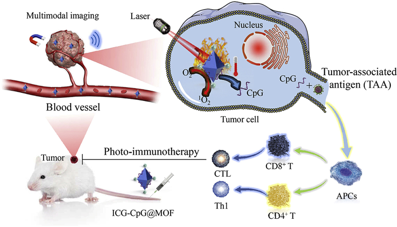
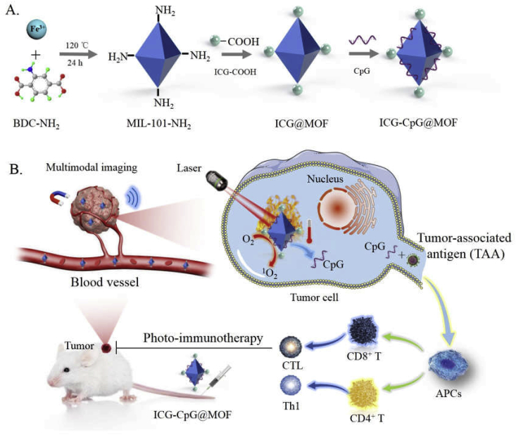
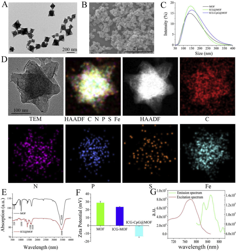
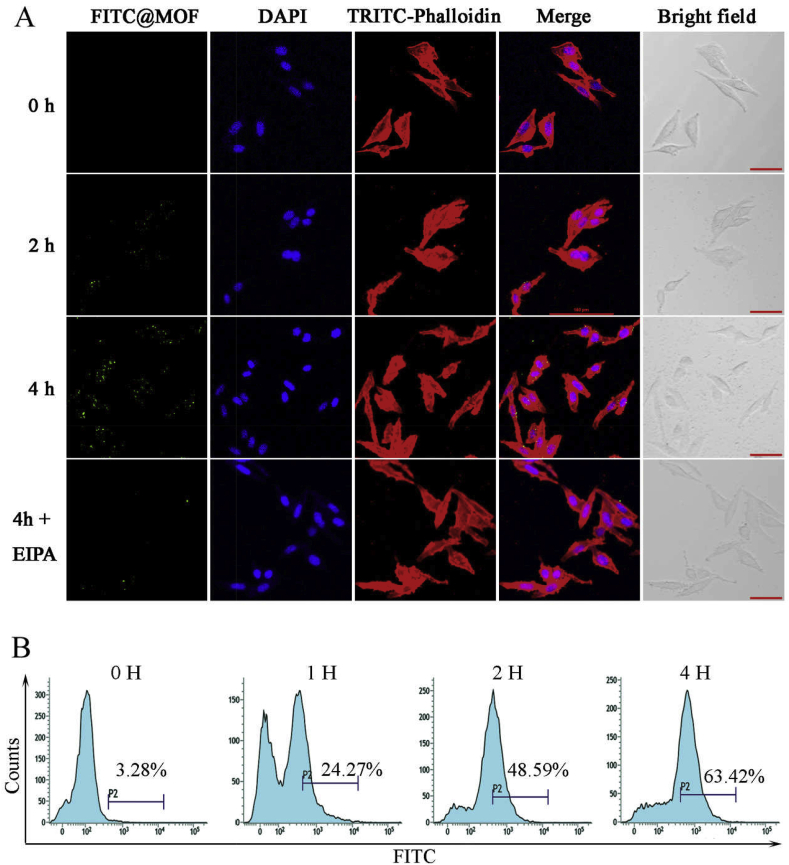
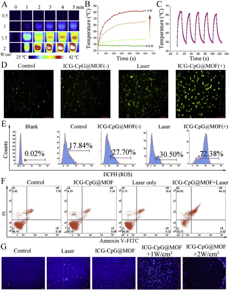
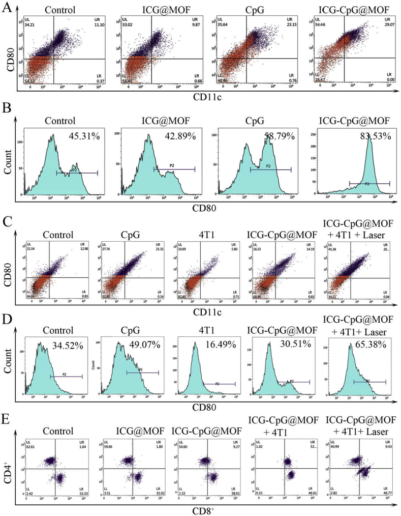
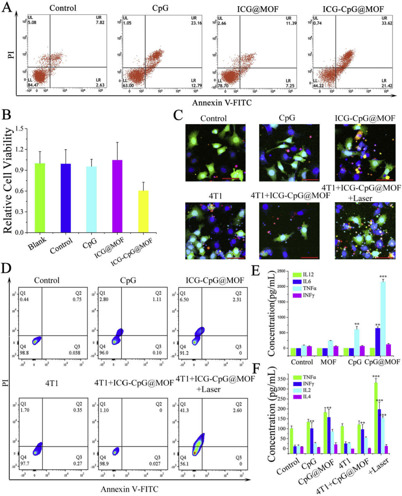
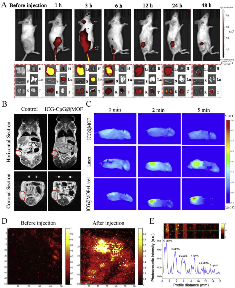
Immunotherapy assays using immunoadjuvants and tumor antigens could greatly increase the survival rates of patients with malignant tumors. As effective carriers, metal-organic frameworks (MOFs) have been widely utilized in cancer therapy due to their remarkable histocompatibility and low toxicity. Herein, we constructed a multimodal imaging-guided synergistic cancer photoimmunotherapy by employing a specific MOF (MIL101-NH) as the core carrier; the MOF was dual-dressed with photoacoustic and fluorescent signal donors (indocyanine green, ICG) and immune adjuvants (cytosine-phosphate-guanine sequence, CpG) and named ICG-CpG@MOF. This nanocarrier could passively target the tumor site through the EPR effect and achieve multimodal imaging (fluorescence, photoacoustic, photothermal and magnetic resonance imaging) of the tumor. Synergistic cancer photoimmunotherapy was achieved via simultaneous photodynamic and photothermal methods with 808 nm laser irradiation. ICG-CpG@MOF achieved the GSH-controlled release of immunoadjuvant into the tumor microenvironment. Furthermore, the released tumor-associated antigen along with CpG could induce the transformation of tumor cells from cold to hot by activating the immune system, which significantly enhanced tumor cytotoxicity and achieved high cure rates with minimal side-effects. This strategy utilizing multimodal imaging and synergistic cancer photoimmunotherapy provides a promising approach for the diagnosis and treatment of cancer.
使用免疫佐剂和肿瘤抗原的免疫疗法检测可大幅提高恶性肿瘤患者的生存率。金属有机框架(MOF)作为有效的载体,因其卓越的组织相容性和低毒性,已在癌症治疗中得到广泛应用。在此,我们构建了一种多模态成像引导的协同癌症光免疫疗法,采用特定的MOF(MIL101-NH)作为核心载体;该MOF用光声和荧光信号供体(吲哚菁绿,ICG)以及免疫佐剂(胞嘧啶-磷酸-鸟嘌呤序列,CpG)进行双重修饰,命名为ICG-CpG@MOF。这种纳米载体可通过EPR效应被动靶向肿瘤部位,实现肿瘤的多模态成像(荧光、光声、光热和磁共振成像)。通过808 nm激光照射,采用光动力和光热方法同时实现了协同癌症光免疫疗法。ICG-CpG@MOF实现了免疫佐剂在肿瘤微环境中的谷胱甘肽控制释放。此外,释放的肿瘤相关抗原与CpG一起可通过激活免疫系统诱导肿瘤细胞从冷肿瘤转变为热肿瘤,显著增强肿瘤细胞毒性,并以最小的副作用实现高治愈率。这种利用多模态成像和协同癌症光免疫疗法的策略为癌症的诊断和治疗提供了一种有前景的方法。