Scott-Ritchey Research Center, College of Veterinary Medicine, Auburn University , Auburn, AL, USA.
Department of Anatomy, Physiology & Pharmacology, College of Veterinary Medicine, Auburn University , Auburn, AL, USA.
Adipocyte. 2020 Dec;9(1):567-575. doi: 10.1080/21623945.2020.1823139.
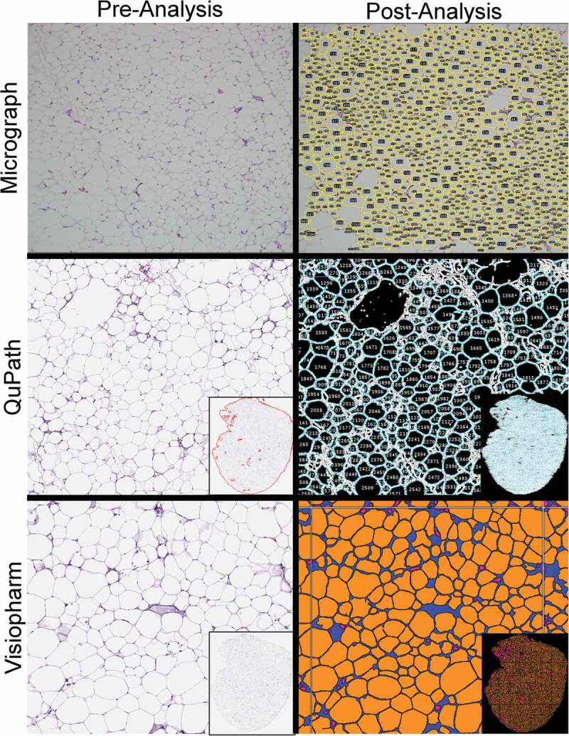
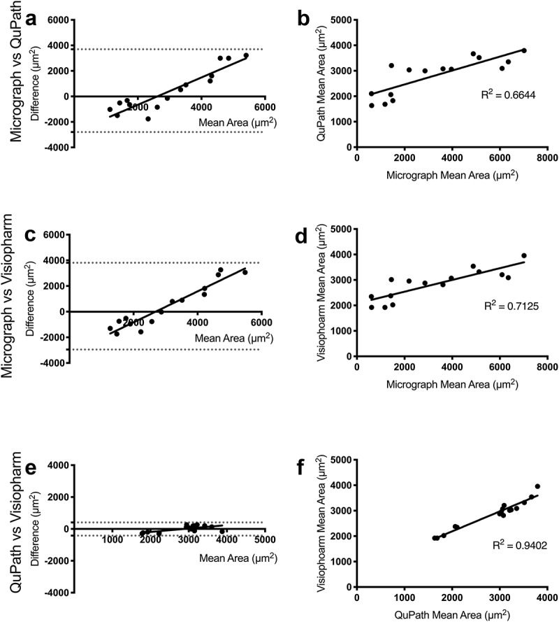
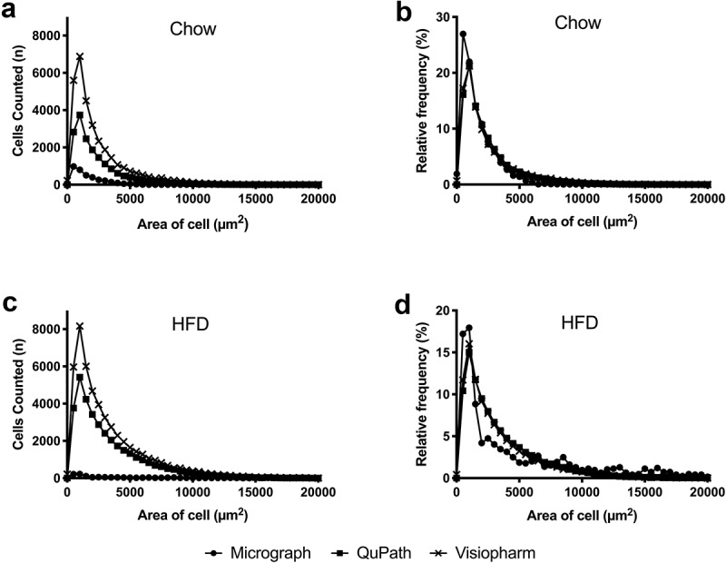
The distinction between biological processes of adipose tissue expansion is crucial to understanding metabolic derangements, but a robust method for quantifying adipocyte size has yet to be standardized. Here, we compared three methods for histological analysis : one conventional approach using individual micrographs acquired by digital camera, and two with whole-slide image analysis pipelines involving proprietary (Visiopharm) and open-source software (QuPath with a novel ImageJ plugin). We found that micrograph analysis identified 10-40 times fewer adipocytes than whole-slide methods, and this small sample size resulted in high variances that could lead to statistical errors. The agreement of the micrograph method to measure adipocyte area with each of the two whole-slide methods was substantially less (R of 0.6644 and 0.7125) than between the two whole-slide methods (R of 0.9402). These inconsistencies were more pronounced in samples from high-fat diet fed mice. While the use of proprietary software resulted in the highest adipocyte count, the lower cost, ease of use, and minimal variances of the open-source software provided a distinct advantage for measuring the number and size of adipocytes. In conclusion, we recommend whole-slide image analysis methods to consistently measure adipocyte area and avoid unintentional errors due to small sample sizes.
区分脂肪组织扩张的生物学过程对于理解代谢紊乱至关重要,但尚未标准化一种用于定量脂肪细胞大小的稳健方法。在这里,我们比较了三种组织学分析方法:一种是使用数码相机获取单个显微照片的传统方法,另外两种是使用具有专有(Visiopharm)和开源软件(带有新型 ImageJ 插件的 QuPath)的全幻灯片图像分析管道。我们发现,与全幻灯片方法相比,显微照片分析方法识别的脂肪细胞数量少 10-40 倍,这种小样本量导致方差很高,可能导致统计误差。显微照片方法与两种全幻灯片方法之一测量脂肪细胞面积的一致性(R 值分别为 0.6644 和 0.7125)明显低于两种全幻灯片方法之间的一致性(R 值为 0.9402)。在高脂肪饮食喂养的小鼠样本中,这些不一致性更为明显。虽然专有软件的使用导致了最高的脂肪细胞计数,但开源软件的低成本、易用性和最小方差为测量脂肪细胞数量和大小提供了明显的优势。总之,我们建议使用全幻灯片图像分析方法来一致地测量脂肪细胞面积,并避免由于样本量小而导致的无意误差。