Löfdahl Anna, Jern Andreas, Flyman Samuel, Kåredal Monica, Karlsson Hanna L, Larsson-Callerfelt Anna-Karin
Unit of Lung Biology, Department of Experimental Medical Sciences, Lund University, SE-221 00 Lund, Sweden.
Division of Occupational and Environmental Medicine, Lund University, SE-221 00 Lund, Sweden.
Nanomaterials (Basel). 2020 Sep 18;10(9):1868. doi: 10.3390/nano10091868.
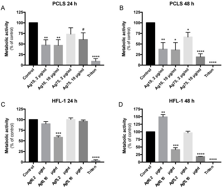
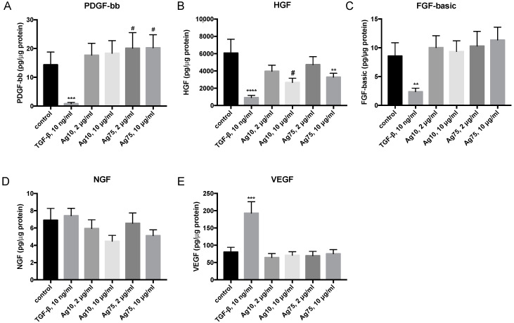
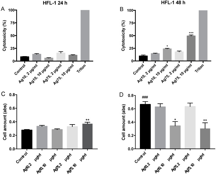
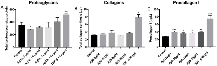
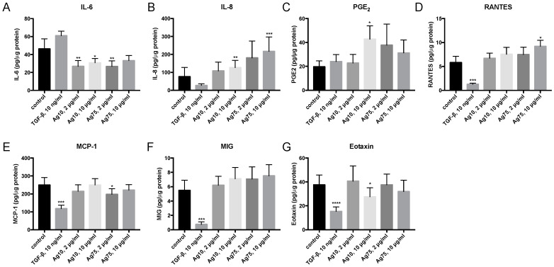
Silver nanoparticles (AgNPs) are commonly used in commercial and medical applications. However, AgNPs may induce toxicity, extracellular matrix (ECM) changes and inflammatory responses. Fibroblasts are key players in remodeling processes and major producers of the ECM. The aims of this study were to explore the effect of AgNPs on cell viability, both ex vivo in murine precision cut lung slices (PCLS) and in vitro in human lung fibroblasts (HFL-1), and immunomodulatory responses in fibroblasts. PCLS and HFL-1 were exposed to AgNPs with different sizes, 10 nm and 75 nm, at concentrations 2 µg/mL and 10 μg/mL. Changes in synthesis of ECM proteins, growth factors and cytokines were analyzed in HFL-1. Ag10 and Ag75 affected cell viability, with significantly reduced metabolic activities at 10 μg/mL in both PCLS and HFL-1 after 48 h. AgNPs significantly increased procollagen I synthesis and release of IL-8, prostaglandin E, RANTES and eotaxin, whereas reduced IL-6 release was observed in HFL-1 after 72 h. Our data indicate toxic effects of AgNP exposure on cell viability ex vivo and in vitro with altered procollagen and proinflammatory cytokine secretion in fibroblasts over time. Hence, careful characterizations of AgNPs are of importance, and future studies should include timepoints beyond 24 h.
银纳米颗粒(AgNPs)常用于商业和医学应用。然而,AgNPs可能会诱导毒性、细胞外基质(ECM)变化和炎症反应。成纤维细胞是重塑过程中的关键参与者和ECM的主要产生者。本研究的目的是探讨AgNPs对细胞活力的影响,包括在小鼠精密切割肺切片(PCLS)中的离体实验以及在人肺成纤维细胞(HFL-1)中的体外实验,以及对成纤维细胞免疫调节反应的影响。将PCLS和HFL-1分别暴露于不同尺寸(10 nm和75 nm)、浓度为2 µg/mL和10 μg/mL的AgNPs。分析了HFL-1中ECM蛋白、生长因子和细胞因子合成的变化。Ag10和Ag75影响细胞活力,48小时后,在PCLS和HFL-1中,10 μg/mL浓度下的代谢活性均显著降低。AgNPs显著增加了I型前胶原的合成以及IL-8、前列腺素E、调节活化正常T细胞表达和分泌的趋化因子(RANTES)和嗜酸性粒细胞趋化因子的释放,而72小时后在HFL-1中观察到IL-6释放减少。我们的数据表明,暴露于AgNPs会对离体和体外细胞活力产生毒性作用,随着时间的推移,成纤维细胞中的前胶原和促炎细胞因子分泌会发生改变。因此,对AgNPs进行仔细的表征非常重要,未来的研究应包括超过24小时的时间点。