• 文献检索
  • 文档翻译
  • 深度研究
  • 学术资讯
  • Suppr Zotero 插件Zotero 插件
  • 邀请有礼
  • 套餐&价格
  • 历史记录
应用&插件
Suppr Zotero 插件Zotero 插件浏览器插件Mac 客户端Windows 客户端微信小程序
定价
高级版会员购买积分包购买API积分包
服务
文献检索文档翻译深度研究API 文档MCP 服务
关于我们
关于 Suppr公司介绍联系我们用户协议隐私条款
关注我们

Suppr 超能文献

核心技术专利:CN118964589B侵权必究
粤ICP备2023148730 号-1Suppr @ 2026

文献检索

告别复杂PubMed语法,用中文像聊天一样搜索,搜遍4000万医学文献。AI智能推荐,让科研检索更轻松。

立即免费搜索

文件翻译

保留排版,准确专业,支持PDF/Word/PPT等文件格式,支持 12+语言互译。

免费翻译文档

深度研究

AI帮你快速写综述,25分钟生成高质量综述,智能提取关键信息,辅助科研写作。

立即免费体验

标准及复杂腔内主动脉修复术中的影像融合:融合与否?一项荟萃分析及单中心回顾性队列的补充数据

Image Fusion During Standard and Complex Endovascular Aortic Repair, to Fuse or Not to Fuse? A Meta-analysis and Additional Data From a Single-Center Retrospective Cohort.

作者信息

Doelare Sabrina A N, Smorenburg Stefan P M, van Schaik Theodorus G, Blankensteijn Jan D, Wisselink Willem, Nederhoed Johanna H, Lely Rutger J, Hoksbergen Arjan W J, Yeung Kak Khee

机构信息

Department of Surgery, Amsterdam Cardiovascular Sciences, Amsterdam UMC, Vrije Universiteit, Amsterdam, the Netherlands.

Department of Radiology, Amsterdam Cardiovascular Sciences, Amsterdam UMC, Vrije Universiteit, Amsterdam, the Netherlands.

出版信息

J Endovasc Ther. 2021 Feb;28(1):78-92. doi: 10.1177/1526602820960444. Epub 2020 Sep 23.

DOI:10.1177/1526602820960444
PMID:32964768
原文链接:https://pmc.ncbi.nlm.nih.gov/articles/PMC7816548/
Abstract

PURPOSE

To determine if image fusion will reduce contrast volume, radiation dose, and fluoroscopy and procedure times in standard and complex (fenestrated/branched) endovascular aneurysm repair (EVAR).

MATERIALS AND METHODS

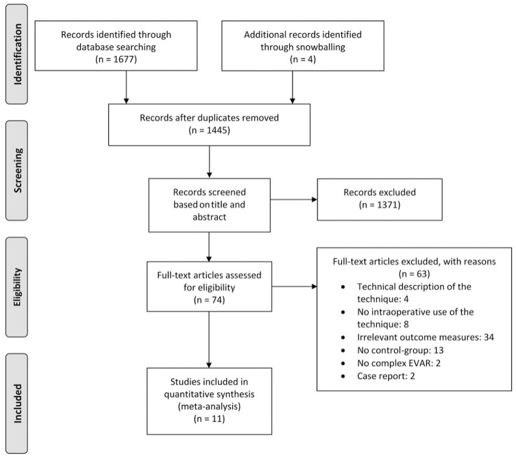
A search of the PubMed, Embase, and Cochrane databases was performed in December 2019 to identify articles describing results of standard and complex EVAR procedures using image fusion compared with a control group. Study selection, data extraction, and assessment of the methodological quality of the included publications were performed by 2 reviewers working independently. Primary outcomes of the pooled analysis were contrast volume, fluoroscopy time, radiation dose, and procedure time. Eleven articles were identified comprising 1547 patients. Data on 140 patients satisfying the study inclusion criteria were added from the authors' center. Mean differences (MDs) are presented with the 95% confidence interval (CI).

RESULTS

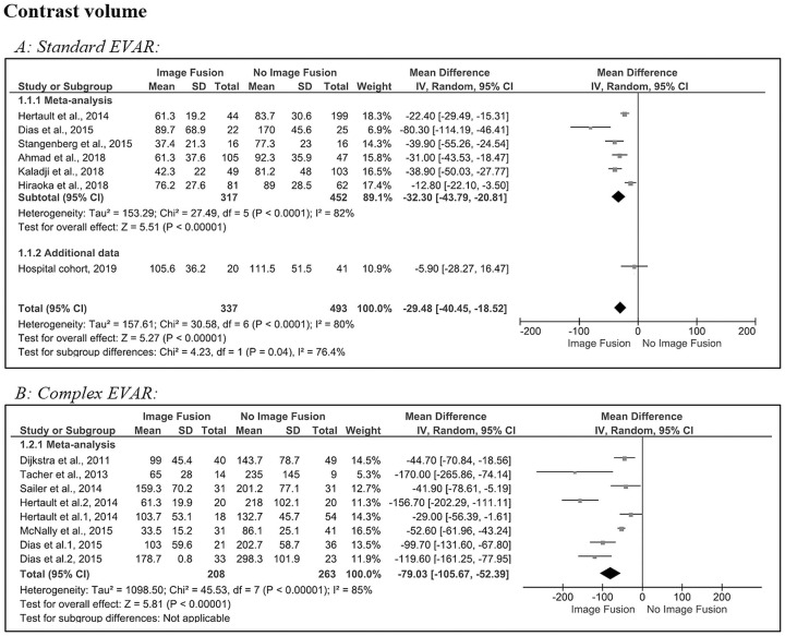
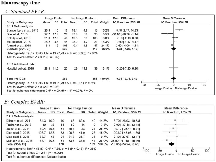
For standard EVAR, contrast volume and procedure time showed a significant reduction with an MD of -29 mL (95% CI -40.5 to -18.5, p<0.001) and -11 minutes (95% CI -21.0 to -1.8, p<0.01), respectively. For complex EVAR, significant reductions in favor of image fusion were found for contrast volume (MD -79 mL, 95% CI -105.7 to -52.4, p<0.001), fluoroscopy time (MD -14 minutes, 95% CI -24.2 to -3.5, p<0.001), and procedure time (MD -52 minutes, 95% CI -75.7 to -27.9, p<0.001).

CONCLUSION

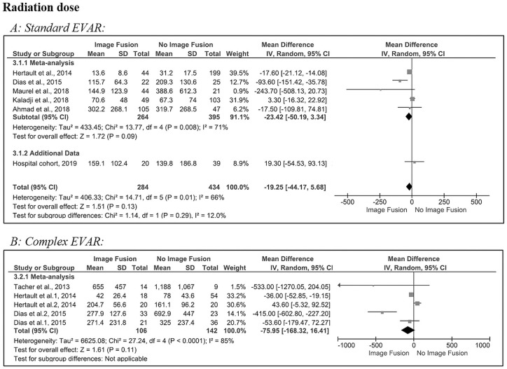
The results of this meta-analysis confirm that image fusion significantly reduces contrast volume, fluoroscopy time, and procedure time in complex EVAR but only contrast volume and procedure time for standard EVAR. Though a reduction was suggested, the radiation dose was not significantly affected by the use of fusion imaging in either standard or complex EVAR.

摘要

目的

确定在标准及复杂(开窗/分支)血管内动脉瘤修复术(EVAR)中,图像融合是否会减少造影剂用量、辐射剂量、透视时间及手术时间。

材料与方法

2019年12月对PubMed、Embase和Cochrane数据库进行检索,以识别描述使用图像融合的标准及复杂EVAR手术结果并与对照组比较的文章。由两名独立工作的审阅者进行研究选择、数据提取及对纳入出版物的方法学质量评估。汇总分析的主要结局为造影剂用量、透视时间、辐射剂量及手术时间。共识别出11篇文章,包含1547例患者。从作者所在中心补充了140例符合研究纳入标准患者的数据。给出平均差值(MD)及95%置信区间(CI)。

结果

对于标准EVAR,造影剂用量和手术时间显著减少,MD分别为-29 mL(95%CI -40.5至-18.5,p<0.001)和-11分钟(95%CI -21.0至-1.8,p<0.01)。对于复杂EVAR,造影剂用量(MD -79 mL,95%CI -105.7至-52.4,p<0.001)、透视时间(MD -14分钟,95%CI -24.2至-3.5,p<0.001)和手术时间(MD -52分钟,95%CI -75.7至-27.9,p<0.001)均显著减少且有利于图像融合。

结论

该荟萃分析结果证实,图像融合在复杂EVAR中显著减少造影剂用量、透视时间和手术时间,但在标准EVAR中仅减少造影剂用量和手术时间。尽管提示辐射剂量有所降低,但在标准或复杂EVAR中,融合成像的使用对辐射剂量均无显著影响。

https://cdn.ncbi.nlm.nih.gov/pmc/blobs/a20a/7816548/567cccbae61e/10.1177_1526602820960444-fig5.jpg
https://cdn.ncbi.nlm.nih.gov/pmc/blobs/a20a/7816548/9997dc7efc40/10.1177_1526602820960444-fig1.jpg
https://cdn.ncbi.nlm.nih.gov/pmc/blobs/a20a/7816548/ea27880d8801/10.1177_1526602820960444-fig2.jpg
https://cdn.ncbi.nlm.nih.gov/pmc/blobs/a20a/7816548/a9e71ad8d834/10.1177_1526602820960444-fig3.jpg
https://cdn.ncbi.nlm.nih.gov/pmc/blobs/a20a/7816548/c80f528b37aa/10.1177_1526602820960444-fig4.jpg
https://cdn.ncbi.nlm.nih.gov/pmc/blobs/a20a/7816548/567cccbae61e/10.1177_1526602820960444-fig5.jpg

相似文献

1
Image Fusion During Standard and Complex Endovascular Aortic Repair, to Fuse or Not to Fuse? A Meta-analysis and Additional Data From a Single-Center Retrospective Cohort.标准及复杂腔内主动脉修复术中的影像融合:融合与否?一项荟萃分析及单中心回顾性队列的补充数据
J Endovasc Ther. 2021 Feb;28(1):78-92. doi: 10.1177/1526602820960444. Epub 2020 Sep 23.
2
Pros and Cons of 3D Image Fusion in Endovascular Aortic Repair: A Systematic Review and Meta-analysis.血管内主动脉修复中3D图像融合的利弊:系统评价与Meta分析
J Endovasc Ther. 2017 Aug;24(4):595-603. doi: 10.1177/1526602817708196. Epub 2017 May 9.
3
The effects of combining fusion imaging, low-frequency pulsed fluoroscopy, and low-concentration contrast agent during endovascular aneurysm repair.血管内动脉瘤修复术中融合成像、低频脉冲荧光透视和低浓度造影剂联合应用的效果。
J Vasc Surg. 2016 May;63(5):1147-55. doi: 10.1016/j.jvs.2015.11.033. Epub 2016 Jan 6.
4
Single-center experience with complex abdominal aortic aneurysms treated by open or endovascular repair using fenestrated/branched endografts.单一中心使用开窗/分支腔内移植物治疗开放或血管内修复复杂腹主动脉瘤的经验。
J Vasc Surg. 2018 Aug;68(2):337-347. doi: 10.1016/j.jvs.2017.11.093. Epub 2018 Mar 3.
5
A prospective observational trial of fusion imaging in infrarenal aneurysms.一项关于肾下动脉瘤融合成像的前瞻性观察性试验。
J Vasc Surg. 2018 Dec;68(6):1706-1713.e1. doi: 10.1016/j.jvs.2018.04.015. Epub 2018 May 24.
6
Dual fluoroscopy with live-image digital zooming significantly reduces patient and operating staff radiation during fenestrated-branched endovascular aortic aneurysm repair.在开窗型分支血管腔内主动脉瘤修复术中,采用实时影像数字变焦的双透视技术可显著降低患者和手术人员的辐射剂量。
J Vasc Surg. 2021 Feb;73(2):601-607. doi: 10.1016/j.jvs.2020.05.031. Epub 2020 May 27.
7
Impact of hybrid rooms with image fusion on radiation exposure during endovascular aortic repair.具有图像融合功能的混合手术室对血管腔内主动脉修复术中辐射暴露的影响。
Eur J Vasc Endovasc Surg. 2014 Oct;48(4):382-90. doi: 10.1016/j.ejvs.2014.05.026. Epub 2014 Jul 17.
8
Monoplane versus biplane fluoroscopy in patients undergoing fenestrated/branched endovascular aortic repair.单机透视与双翼透视在接受开窗/分支腔内主动脉修复的患者中的应用比较。
J Vasc Surg. 2023 May;77(5):1359-1366.e2. doi: 10.1016/j.jvs.2022.12.037. Epub 2022 Dec 29.
9
Three-dimensional fusion computed tomography decreases radiation exposure, procedure time, and contrast use during fenestrated endovascular aortic repair.三维融合计算机断层扫描可减少开窗式血管内主动脉修复术中的辐射暴露、手术时间和造影剂用量。
J Vasc Surg. 2015 Feb;61(2):309-16. doi: 10.1016/j.jvs.2014.07.097. Epub 2014 Aug 28.
10
Dose reduction using digital fluoroscopy versus digital subtraction angiography in endovascular aneurysm repair: A prospective randomized trial.数字减影血管造影与数字荧光透视在血管内动脉瘤修复术中的剂量减少:一项前瞻性随机试验。
J Vasc Surg. 2020 Dec;72(6):1938-1945. doi: 10.1016/j.jvs.2020.02.050. Epub 2020 Apr 8.

引用本文的文献

1
Vessel-based CTA-image to spatial anatomy registration using tracked catheter position data: preclinical evaluation of in vivo accuracy.基于血管的 CTA 图像与基于跟踪导管位置数据的空间解剖配准:体内准确性的临床前评估。
Eur Radiol Exp. 2024 Aug 28;8(1):99. doi: 10.1186/s41747-024-00499-1.
2
Aortic roadmapping during EVAR: a combined FEM-EM tracking feasibility study.腹主动脉腔内修复术中的主动脉路径图:一种联合 FEM-EM 跟踪的可行性研究。
Int J Comput Assist Radiol Surg. 2024 Nov;19(11):2239-2247. doi: 10.1007/s11548-024-03187-y. Epub 2024 Jun 2.
3
Image fusion guidance for left subclavian artery in situ fenestration during thoracic endovascular repair.

本文引用的文献

1
Performance of a feature-based algorithm for 3D-3D registration of CT angiography to cone-beam CT for endovascular repair of complex abdominal aortic aneurysms.一种基于特征的算法用于将CT血管造影与锥形束CT进行三维-三维配准以用于复杂腹主动脉瘤血管内修复的性能。
BMC Med Imaging. 2018 Nov 8;18(1):42. doi: 10.1186/s12880-018-0285-1.
2
Fusion Imaging for EVAR with Mobile C-arm.使用移动C型臂进行腔内隔绝术的融合成像
Ann Vasc Surg. 2019 Feb;55:166-174. doi: 10.1016/j.avsg.2018.06.006. Epub 2018 Aug 6.
3
A prospective observational trial of fusion imaging in infrarenal aneurysms.
图像融合引导技术在胸主动脉腔内修复术中左锁骨下动脉原位开窗的应用。
J Cardiothorac Surg. 2024 Feb 22;19(1):104. doi: 10.1186/s13019-024-02561-w.
4
"Redo" 2D-3D Fusion Technique during Endovascular Redo Aortic Repair.血管腔内再次主动脉修复术中的“重复”二维-三维融合技术
Diagnostics (Basel). 2023 Feb 8;13(4):635. doi: 10.3390/diagnostics13040635.
5
Automated image fusion during endovascular aneurysm repair: a feasibility and accuracy study.血管内动脉瘤修复术中的自动图像融合:一项可行性和准确性研究。
Int J Comput Assist Radiol Surg. 2023 Aug;18(8):1533-1541. doi: 10.1007/s11548-023-02832-2. Epub 2023 Jan 31.
6
Use of surgical augmented intelligence maps can reduce radiation and improve safety in the endovascular treatment of complex aortic aneurysms.使用手术增强智能图谱可以降低腔内治疗复杂主动脉瘤的辐射剂量并提高安全性。
J Vasc Surg. 2023 Apr;77(4):982-990.e2. doi: 10.1016/j.jvs.2022.12.033. Epub 2022 Dec 26.
7
The Current Era of Endovascular Aortic Interventions and What the Future Holds.当前的血管内主动脉介入治疗时代及未来展望。
J Clin Med. 2022 Oct 6;11(19):5900. doi: 10.3390/jcm11195900.
8
Deep Learning-Based Intraoperative Stent Graft Segmentation on Completion Digital Subtraction Angiography During Endovascular Aneurysm Repair.基于深度学习的血管内动脉瘤修复术完成数字减影血管造影术中支架移植物的自动分割。
J Endovasc Ther. 2023 Dec;30(6):822-827. doi: 10.1177/15266028221105840. Epub 2022 Jul 9.
一项关于肾下动脉瘤融合成像的前瞻性观察性试验。
J Vasc Surg. 2018 Dec;68(6):1706-1713.e1. doi: 10.1016/j.jvs.2018.04.015. Epub 2018 May 24.
4
Procedure and step-based analysis of the occupational radiation dose during endovascular aneurysm repair in the hybrid operating room.在杂交手术室进行血管内动脉瘤修复时的职业辐射剂量的基于程序和步骤的分析。
J Vasc Surg. 2018 Jun;67(6):1881-1890. doi: 10.1016/j.jvs.2017.09.043.
5
Feasibility of three-dimensional fusion imaging with multimodality roadmap system during endovascular aortic repair.多模态路标系统三维融合成像在腹主动脉瘤腔内修复术中的可行性。
J Vasc Surg. 2018 Oct;68(4):1175-1182. doi: 10.1016/j.jvs.2017.12.065. Epub 2018 Mar 31.
6
The 2D-3D Registration Method in Image Fusion Is Accurate and Helps to Reduce the Used Contrast Medium, Radiation, and Procedural Time in Standard EVAR Procedures.图像融合中的二维-三维配准方法准确无误,有助于在标准腔内血管修复术中减少造影剂用量、辐射剂量及手术时间。
Ann Vasc Surg. 2018 Aug;51:177-186. doi: 10.1016/j.avsg.2018.01.098. Epub 2018 Mar 6.
7
Complications and Reinterventions After Fenestrated and Branched EVAR in Patients with Paravisceral and Thoracoabdominal Aneurysms.
Cardiovasc Intervent Radiol. 2018 Jul;41(7):985-997. doi: 10.1007/s00270-018-1917-0. Epub 2018 Mar 6.
8
Computed tomography angiography-fluoroscopy image fusion allows visceral vessel cannulation without angiography during fenestrated endovascular aneurysm repair.计算机断层血管造影-透视图像融合可在血管内开窗动脉瘤修复术中无需血管造影即可进行内脏血管插管。
J Vasc Surg. 2018 Jul;68(1):2-11. doi: 10.1016/j.jvs.2017.11.062. Epub 2018 Feb 1.
9
Pros and Cons of 3D Image Fusion in Endovascular Aortic Repair: A Systematic Review and Meta-analysis.血管内主动脉修复中3D图像融合的利弊:系统评价与Meta分析
J Endovasc Ther. 2017 Aug;24(4):595-603. doi: 10.1177/1526602817708196. Epub 2017 May 9.
10
Radiation Awareness for Endovascular Abdominal Aortic Aneurysm Repair in the Hybrid Operating Room. An Instant Patient Risk Chart for Daily Practice.杂交手术室中腹主动脉瘤腔内修复术的辐射防护。日常实践中的即时患者风险图表。
J Endovasc Ther. 2017 Jun;24(3):425-434. doi: 10.1177/1526602817697188. Epub 2017 Apr 10.