Molecular Cardiology, Department of Cardiology and Angiology, Hannover Medical School, Carl-Neuberg Str. 1, 30625, Hannover, Germany.
Department of Hematology, Hemostasis, Oncology and Stem Cell Transplantation, Hannover Medical School, Carl-Neuberg-Str.1, 30625, Hannover, Germany.
Basic Res Cardiol. 2020 Sep 25;115(6):62. doi: 10.1007/s00395-020-00821-z.
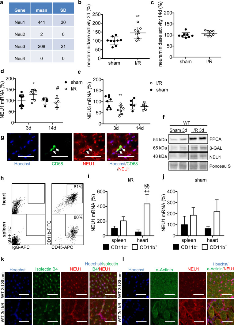
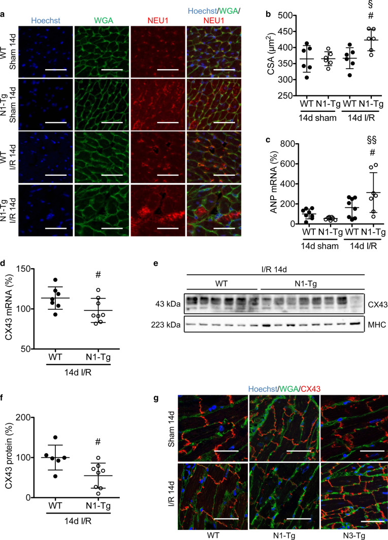
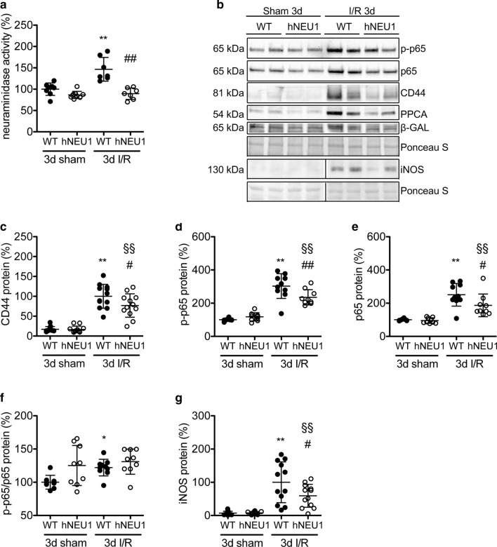
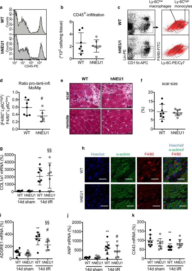
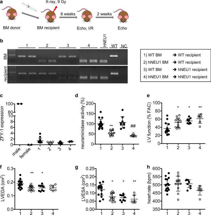
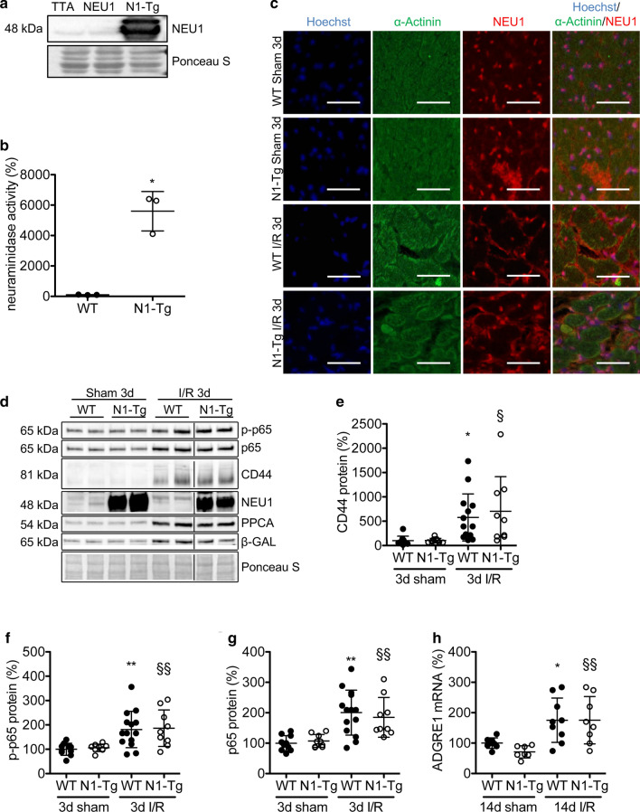
Neuraminidase (NEU)1 forms a multienzyme complex with beta-galactosidase (β-GAL) and protective-protein/cathepsin (PPC) A, which cleaves sialic-acids from cell surface glycoconjugates. We investigated the role of NEU1 in the myocardium after ischemia/reperfusion (I/R). Three days after inducing I/R, left ventricles (LV) of male mice (3 months-old) displayed upregulated neuraminidase activity and increased NEU1, β-GAL and PPCA expression. Mice hypomorphic for neu1 (hNEU1) had less neuraminidase activity, fewer pro-inflammatory (LinCD11bF4/80Ly-6C), and more anti-inflammatory macrophages (LinCD11bF4/80Ly-6C) 3 days after I/R, and less LV dysfunction 14 days after I/R. WT mice transplanted with hNEU1-bone marrow (BM) and hNEU1 mice with WT-BM showed significantly better LV function 14 days after I/R compared with WT mice with WT-BM. Mice with a cardiomyocyte-specific NEU1 overexpression displayed no difference in inflammation 3 days after I/R, but showed increased cardiomyocyte hypertrophy, reduced expression and mislocalization of Connexin-43 in gap junctions, and LV dysfunction despite a similar infarct scar size to WT mice 14 days after I/R. The upregulation of NEU1 after I/R contributes to heart failure by promoting inflammation in invading monocytes/macrophages, enhancing cardiomyocyte hypertrophy, and impairing gap junction function, suggesting that systemic NEU1 inhibition may reduce heart failure after I/R.
神经氨酸酶(NEU)1 与β-半乳糖苷酶(β-GAL)和保护蛋白/组织蛋白酶(PPC)A 形成多酶复合物,从细胞表面糖缀合物上切割唾液酸。我们研究了 NEU1 在缺血/再灌注(I/R)后心肌中的作用。在诱导 I/R 后 3 天,雄性小鼠(3 个月大)的左心室(LV)显示出神经氨酸酶活性上调,NEU1、β-GAL 和 PPC 表达增加。neu1 功能减弱(hNEU1)的小鼠在 I/R 后 3 天具有较少的神经氨酸酶活性、较少的促炎(LinCD11bF4/80Ly-6C)和更多的抗炎巨噬细胞(LinCD11bF4/80Ly-6C),并且在 I/R 后 14 天 LV 功能障碍较少。WT 小鼠接受 hNEU1-骨髓(BM)移植和 hNEU1 小鼠接受 WT-BM 显示出在 I/R 后 14 天与 WT 小鼠接受 WT-BM 相比,LV 功能明显更好。在 I/R 后 3 天,具有心肌细胞特异性 NEU1 过表达的小鼠在炎症方面没有差异,但表现出心肌细胞肥大增加、间隙连接中 Connexin-43 的表达和定位减少,以及 LV 功能障碍,尽管梗死疤痕大小与 WT 小鼠相似在 I/R 后 14 天。I/R 后 NEU1 的上调通过促进浸润单核细胞/巨噬细胞中的炎症、增强心肌细胞肥大以及损害间隙连接功能,导致心力衰竭,提示全身 NEU1 抑制可能减少 I/R 后的心力衰竭。