Ishikawa Koji, Nakao Yusuke, Oguchi Fumihiko, Toyone Tomoaki, Sano Shigeo
Sanraku Hospital, Chiyoda, Tokyo, Japan.
Showa University, Shinagawa, Tokyo, Japan.
Global Spine J. 2021 Oct;11(8):1230-1237. doi: 10.1177/2192568220944169. Epub 2020 Sep 29.
Retrospective cohort study.
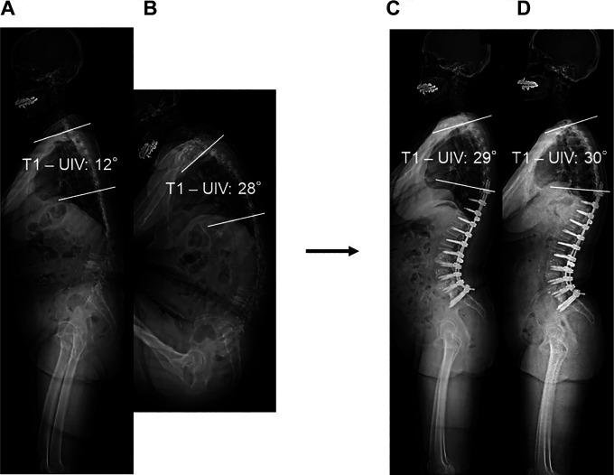
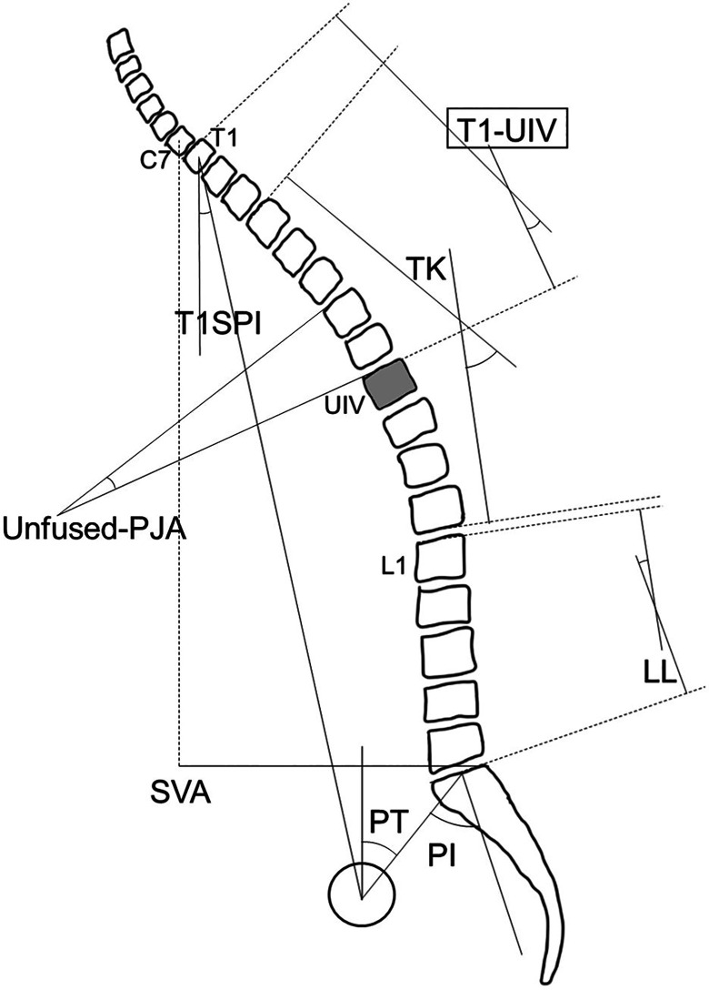
Analysis of postoperative sagittal alignment of the unfused spine is lacking in patients with adult spinal deformity (ASD). The present study aims to evaluate the efficacy of the whole spine full-flexion lateral radiograph to predict the reciprocal change of the unfused spine after correction surgery. We hypothesized that the novel parameter (T1-UIV angle: angle between the upper vertebral endplate of the T1 and the upper vertebral endplate of the upper instrumented vertebra) of the preoperative whole spine full-flexion lateral radiograph is similar to that of the postoperative lateral radiograph if the patient has the ideal sagittal alignment.
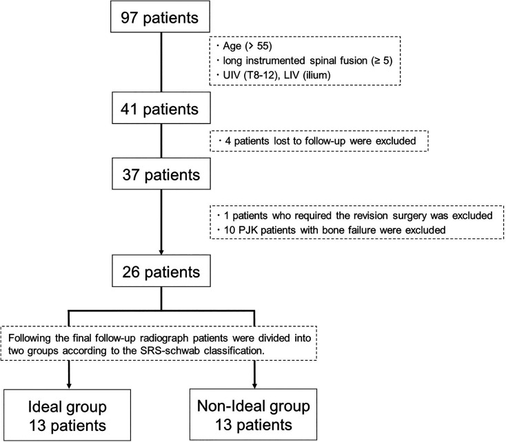
Twenty-six ASD patients who underwent correction surgery with a minimum 2-year follow-up were enrolled and separated into the Ideal and Non-Ideal groups according to the Scoliosis Research Society (SRS)-Schwab classification of the final follow-up radiograph. Radiographic parameters, including T1-UIV of the preoperative whole spine full-flexion lateral radiograph, were obtained.
Thirteen patients were included in the Ideal group and 13 were in Non-Ideal group. Preoperative T1-UIV of the whole spine full-flexion lateral radiograph exhibited significant correlations with the T1-UIV angles of the postoperative and final follow-up radiographs ( = 0.64, < .01, = 0.800 + 8.012, and = 0.69, < .01, = 0.857 + 2.960, respectively). Interestingly, this correlation was stronger for the Ideal group ( = 0.77, < .01, = 1.207 - 1.517, and = 0.89, < .01, = 0.986 + 0.694, respectively).
A novel radiographic strategy (T1-UIV of preoperative the whole spine full-flexion lateral radiograph) could estimate the postoperative alignment of the unfused spine correctly.
回顾性队列研究。
成人脊柱畸形(ASD)患者缺乏对未融合脊柱术后矢状面排列的分析。本研究旨在评估全脊柱全屈侧位X线片预测矫正手术后未融合脊柱相互变化的有效性。我们假设,如果患者矢状面排列理想,术前全脊柱全屈侧位X线片的新参数(T1 - UIV角:T1上终板与上固定椎上终板之间的夹角)与术后侧位X线片的参数相似。
纳入26例接受矫正手术且至少随访2年的ASD患者,并根据最终随访X线片的脊柱侧弯研究学会(SRS)-施瓦布分类分为理想组和非理想组。获取包括术前全脊柱全屈侧位X线片的T1 - UIV在内的影像学参数。
理想组纳入13例患者,非理想组纳入13例患者。术前全脊柱全屈侧位X线片的T1 - UIV与术后及最终随访X线片的T1 - UIV角均呈显著相关(分别为r = 0.64,P <.01,y = 0.800x + 8.012,以及r = 0.69,P <.01,y = 0.857x + 2.960)。有趣的是,这种相关性在理想组中更强(分别为r = 0.77,P <.01,y = 1.207x - 1.517,以及r = 0.89,P <.01,y = 0.986x + 0.694)。
一种新的影像学策略(术前全脊柱全屈侧位X线片的T1 - UIV)能够正确估计未融合脊柱的术后排列情况。