Suurs Frans V, Qiu Si-Qi, Yim Joshua J, Schröder Carolien P, Timmer-Bosscha Hetty, Bensen Eric S, Santini John T, de Vries Elisabeth G E, Bogyo Matthew, van Dam Gooitzen M
Department of Medical Oncology, University of Groningen, University Medical Center Groningen, Groningen, The Netherlands.
Diagnosis and Treatment Center of Breast Diseases, Affiliated Shantou Hospital, Sun Yat-Sen University, Shantou, China.
EJNMMI Res. 2020 Sep 29;10(1):111. doi: 10.1186/s13550-020-00688-0.
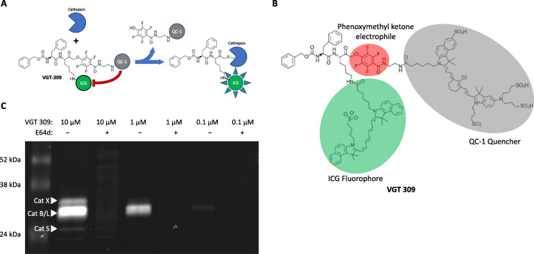
The reoperation rate for breast-conserving surgery is as high as 15-30% due to residual tumor in the surgical cavity after surgery. In vivo tumor-targeted optical molecular imaging may serve as a red-flag technique to improve intraoperative surgical margin assessment and to reduce reoperation rates. Cysteine cathepsins are overexpressed in most solid tumor types, including breast cancer. We developed a cathepsin-targeted, quenched fluorescent activity-based probe, VGT-309, and evaluated whether it could be used for tumor detection and image-guided surgery in syngeneic tumor-bearing mice.
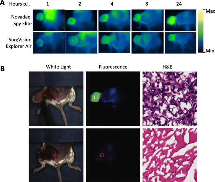
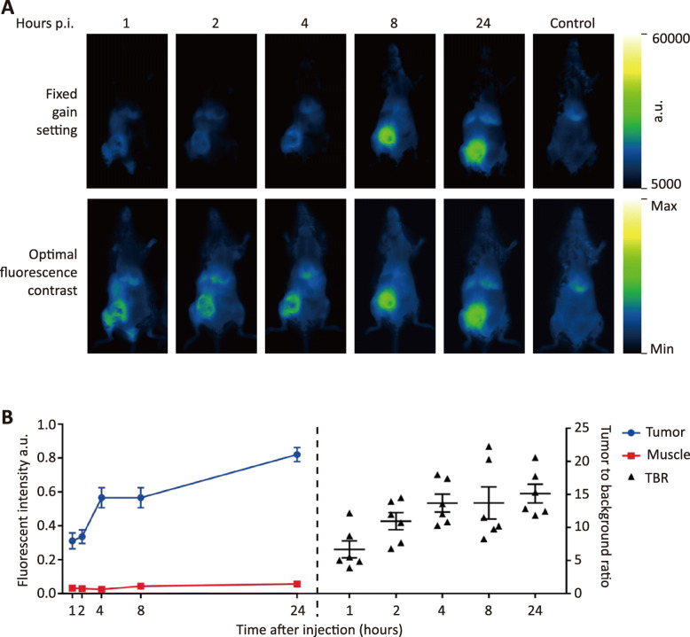
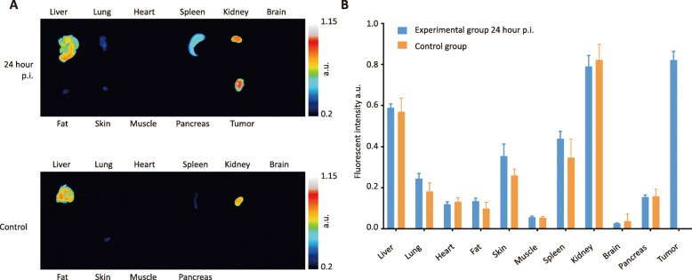
Binding specificity of the developed probe was evaluated in vitro. Next, fluorescent imaging in BALB/c mice bearing a murine breast tumor was performed at different time points after VGT-309 administration. Biodistribution of VGT-309 after 24 h in tumor-bearing mice was compared to control mice. Image-guided surgery was performed at multiple time points tumors with different clinical fluorescent camera systems and followed by ex vivo analysis.
The probe was specifically activated by cathepsins X, B/L, and S. Fluorescent imaging revealed an increased tumor-to-background contrast over time up to 15.1 24 h post probe injection. In addition, VGT-309 delineated tumor tissue during image-guided surgery with different optical fluorescent imaging camera systems.
These results indicate that optical fluorescent molecular imaging using the cathepsin-targeted probe, VGT-309, may improve intraoperative tumor detection, which could translate to more complete tumor resection when coupled with commercially available surgical tools and techniques.
由于保乳手术后手术腔内残留肿瘤,保乳手术的再次手术率高达15%-30%。体内肿瘤靶向光学分子成像可作为一种警示技术,以改善术中手术切缘评估并降低再次手术率。半胱氨酸组织蛋白酶在包括乳腺癌在内的大多数实体瘤类型中过表达。我们开发了一种靶向组织蛋白酶、基于荧光活性淬灭的探针VGT-309,并评估其是否可用于同基因荷瘤小鼠的肿瘤检测和图像引导手术。
在体外评估所开发探针的结合特异性。接下来,在给予VGT-309后的不同时间点,对携带小鼠乳腺肿瘤的BALB/c小鼠进行荧光成像。将荷瘤小鼠在24小时后VGT-309的生物分布与对照小鼠进行比较。使用不同的临床荧光摄像系统在多个时间点对肿瘤进行图像引导手术,然后进行离体分析。
该探针被组织蛋白酶X、B/L和S特异性激活。荧光成像显示,在注射探针后长达24小时内,肿瘤与背景的对比度随时间增加,最高可达15.1。此外,VGT-309在使用不同光学荧光成像摄像系统的图像引导手术中勾勒出肿瘤组织。
这些结果表明,使用靶向组织蛋白酶的探针VGT-309进行光学荧光分子成像可能会改善术中肿瘤检测,当与市售手术工具和技术结合使用时,这可能会实现更完整的肿瘤切除。