Tsubaki Yuma, Akamatsu Go, Shimokawa Natsumi, Katsube Suguru, Takashima Aya, Sasaki Masayuki
Department of Medical Quantum Science, Graduate School of Medical Sciences, Kyushu University, 3-1-1 Maidashi, Higashi-ku, Fukuoka, 812-8582, Japan.
National Institute of Radiological Sciences, National Institutes for Quantum and Radiological Science and Technology (NIRS-QST), 4-9-1 Anagawa, Inage-ku, Chiba, 263-8555, Japan.
EJNMMI Phys. 2020 Sep 29;7(1):59. doi: 10.1186/s40658-020-00329-4.
Quantitative evaluation of amyloid positron emission tomography (PET) with standardized uptake value ratio (SUVR) plays a key role in clinical studies of Alzheimer's disease (AD). We have proposed a PET-only (MR-free) amyloid quantification method, although some commercial software packages are required. The aim of this study was to develop an automated quantification tool for amyloid PET without using commercial software.
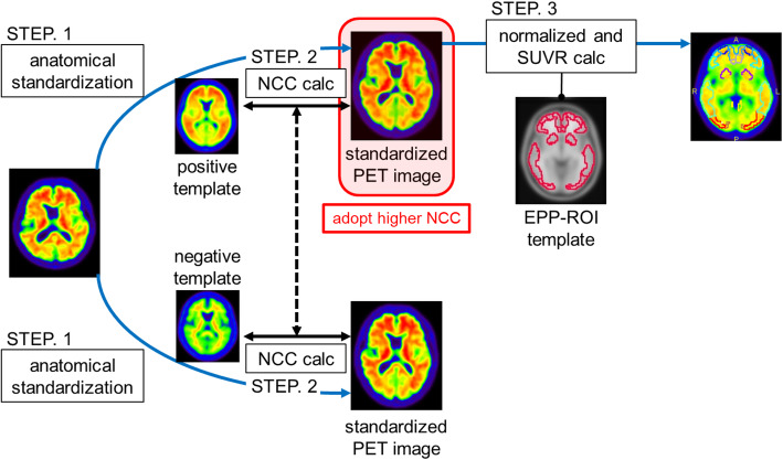
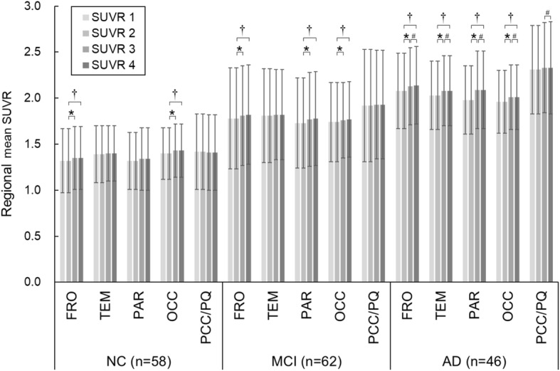
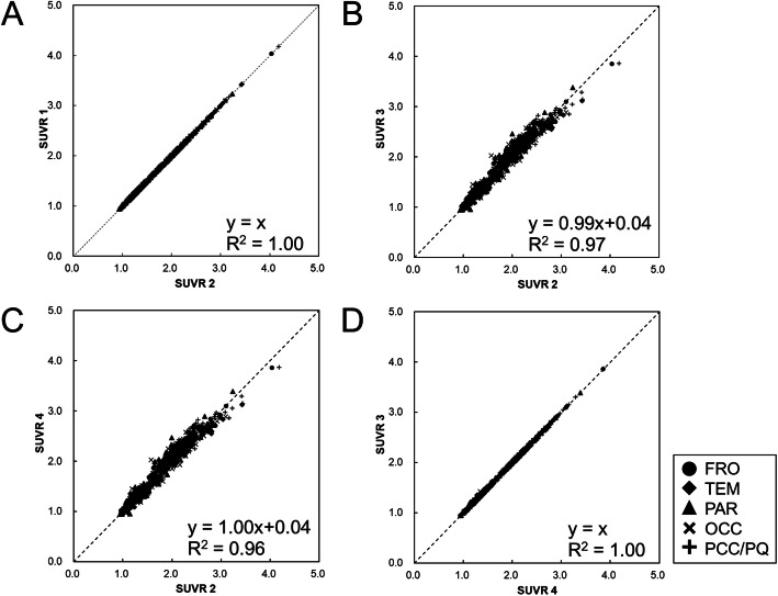
The quantification tool was created by combining four components: (1) anatomical standardization to positive and negative templates using NEUROSTAT stereo.exe; (2) similarity calculation between standardized images and respective templates based on normalized cross-correlation (selection of the image for SUVR measurement); (3) voxel value normalization by the mean value of reference regions (making an SUVR-scaled image); and (4) SUVR calculation based on pre-defined regions of interest (ROIs). We examined 166 subjects who underwent a [C] Pittsburgh compound-B PET scan through the Japanese Alzheimer's Disease Neuroimaging Initiative (J-ADNI) study. SUVRs in five ROIs (frontal lobe, temporal lobe, parietal lobe, occipital lobe, and posterior cingulate cortex and precuneus) were calculated with the cerebellar cortex as the reference region. The SUVRs obtained by our tool were compared with manual step-by-step processing and the conventional PMOD-based method (PMOD Technologies, Switzerland).
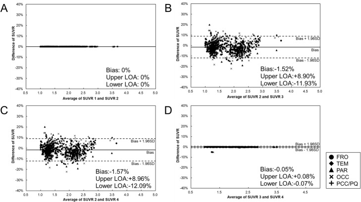
Compared with manual step-by-step processing, our developed automated quantification tool reduced processing time by 85%. The SUVRs obtained by the developed quantification tool were consistent with those obtained by manual processing. Compared with the conventional PMOD-based method, the developed quantification tool provided 1.5% lower SUVR values, on average. We determined that this bias is likely due to the difference in anatomical standardization methods.
We developed an automated quantification tool for amyloid PET images. Using this tool, SUVR values can be quickly measured without individual MRI and without commercial software. This quantification tool may be useful for clinical studies of AD.
采用标准化摄取值比率(SUVR)对淀粉样蛋白正电子发射断层扫描(PET)进行定量评估在阿尔茨海默病(AD)临床研究中起着关键作用。我们已经提出了一种仅使用PET(无需磁共振成像)的淀粉样蛋白定量方法,不过需要一些商业软件包。本研究的目的是开发一种无需使用商业软件的淀粉样蛋白PET自动定量工具。
该定量工具由四个部分组成:(1)使用NEUROSTAT stereo.exe对阳性和阴性模板进行解剖标准化;(2)基于归一化互相关计算标准化图像与各自模板之间的相似度(选择用于SUVR测量的图像);(3)通过参考区域的平均值进行体素值归一化(生成SUVR缩放图像);(4)基于预定义的感兴趣区域(ROI)计算SUVR。我们通过日本阿尔茨海默病神经影像学倡议(J-ADNI)研究检查了166名接受了[C]匹兹堡化合物B PET扫描的受试者。以小脑皮质为参考区域,计算了五个ROI(额叶、颞叶、顶叶、枕叶以及后扣带回皮质和楔前叶)中的SUVR。将我们工具获得的SUVR与手动逐步处理以及传统的基于PMOD的方法(瑞士PMOD Technologies公司)获得的结果进行比较。
与手动逐步处理相比,我们开发的自动定量工具将处理时间减少了85%。开发的定量工具获得的SUVR与手动处理获得的结果一致。与传统的基于PMOD的方法相比,开发的定量工具平均提供的SUVR值低1.5%。我们确定这种偏差可能是由于解剖标准化方法的差异所致。
我们开发了一种用于淀粉样蛋白PET图像的自动定量工具。使用该工具,可以在无需个体磁共振成像且无需商业软件的情况下快速测量SUVR值。这种定量工具可能对AD临床研究有用。