Department of Molecular Pathology, Nara Medical University, 840 Shijo-cho, Kashihara, Nara 634-8521, Japan.
Department of Oral and Maxillofacial Surgery, Nara Medical University, 840 Shijo-cho, Kashihara, Nara 634-8522, Japan.
Int J Mol Sci. 2020 Sep 28;21(19):7149. doi: 10.3390/ijms21197149.
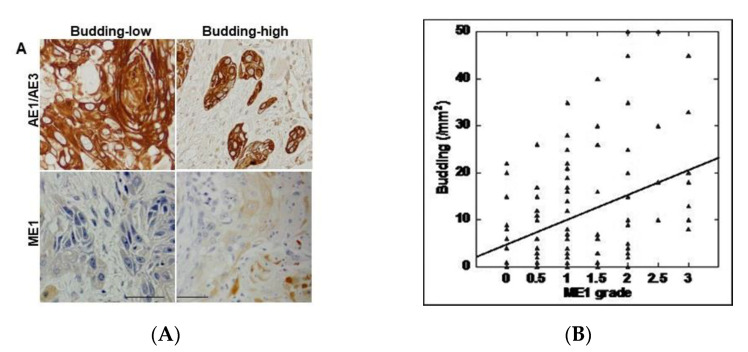
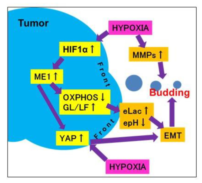
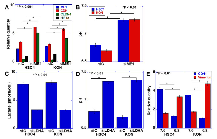
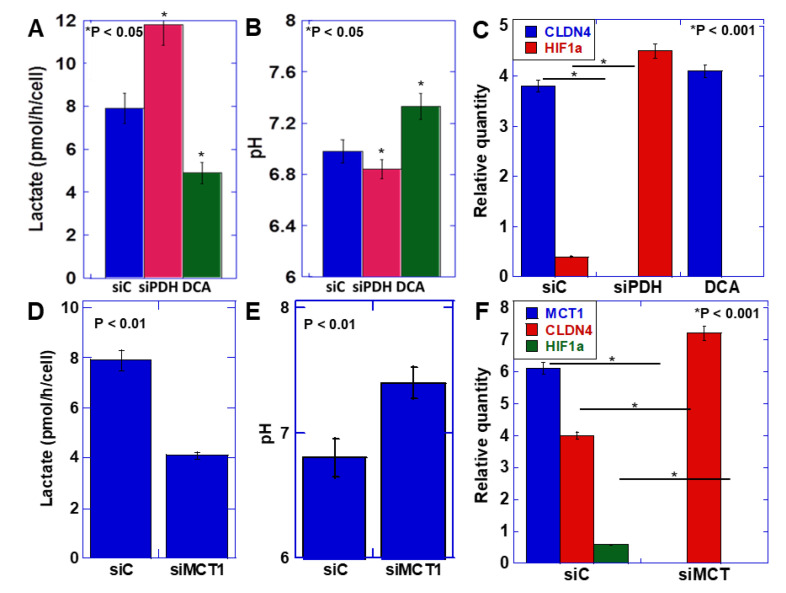
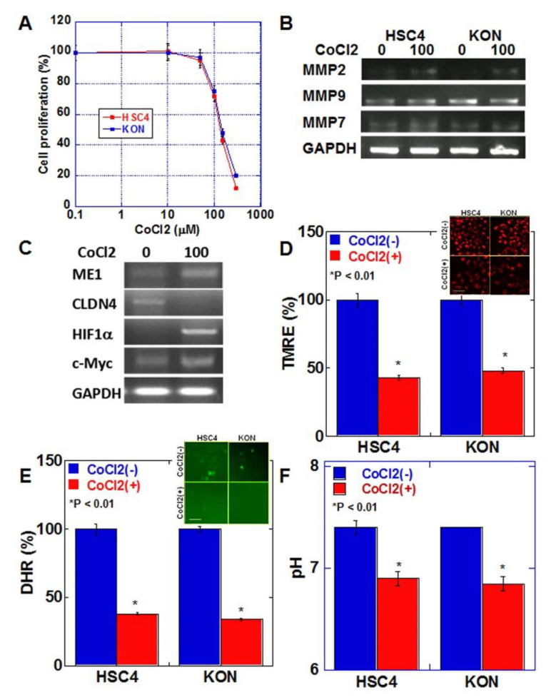
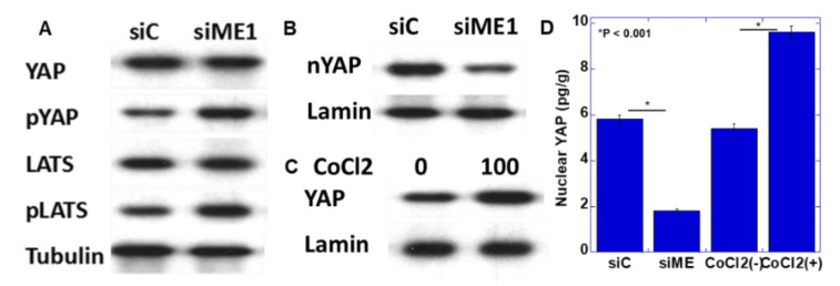
Budding at the tumor invasive front has been correlated with the malignant properties of many cancers. Malic enzyme 1 (ME1) promotes the Warburg effect in cancer cells and induces epithelial-mesenchymal transition (EMT) in oral squamous cell carcinoma (OSCC). Therefore, we investigated the role of ME1 in tumor budding in OSCC. Tumor budding was measured in 96 human OSCCs by immunostaining for an epithelial marker (AE1/AE3), and its expression was compared with that of ME1. A significant correlation was observed between tumor budding and ME1 expression. The correlation increased with the progression of cancer. In human OSCC cells, lactate secretion decreased when lactate fermentation was suppressed by knockdown of ME1 and lactate dehydrogenase A or inhibition of pyruvate dehydrogenase (PDH) kinase. Furthermore, the extracellular pH increased, and the EMT phenotype was suppressed. In contrast, when oxidative phosphorylation was suppressed by PDH knockdown, lactate secretion increased, extracellular pH decreased, and the EMT phenotype was promoted. Induction of chemical hypoxia in OSCC cells by CoCl treatment resulted in increased ME1 expression along with HIF1α expression and promotion of the EMT phenotype. Hypoxic conditions also increased matrix metalloproteinases expression and decreased mitochondrial membrane potential, mitochondrial oxidative stress, and extracellular pH. Furthermore, the hypoxic treatment resulted in the activation of Yes-associated protein (YAP), which was abolished by ME1 knockdown. These findings suggest that cancer cells at the tumor front in hypoxic environments increase their lactate secretion by switching their energy metabolism from oxidative phosphorylation to glycolysis owing to ME1 overexpression, decrease in extracellular pH, and YAP activation. These alterations enhance EMT and the subsequent tumor budding. Tumor budding and ME1 expression are thus considered useful markers of OSCC malignancy, and ME1 is expected to be a relevant target for molecular therapy.
肿瘤侵袭前沿的出芽与许多癌症的恶性特性有关。苹果酸酶 1(ME1)促进癌细胞的瓦博格效应,并诱导口腔鳞状细胞癌(OSCC)中的上皮-间充质转化(EMT)。因此,我们研究了 ME1 在 OSCC 肿瘤出芽中的作用。通过免疫染色上皮标志物(AE1/AE3)测量了 96 个人类 OSCC 中的肿瘤出芽,并将其表达与 ME1 的表达进行了比较。观察到肿瘤出芽与 ME1 表达之间存在显著相关性。该相关性随着癌症的进展而增加。在人类 OSCC 细胞中,当通过敲低 ME1 和乳酸脱氢酶 A 或抑制丙酮酸脱氢酶(PDH)激酶抑制乳酸发酵时,乳酸分泌减少。此外,细胞外 pH 值升高,EMT 表型受到抑制。相反,当通过 PDH 敲低抑制氧化磷酸化时,乳酸分泌增加,细胞外 pH 值降低,EMT 表型得到促进。用 CoCl 处理 OSCC 细胞诱导化学缺氧会导致 ME1 表达增加,同时 HIF1α 表达增加,并促进 EMT 表型。缺氧条件还增加了基质金属蛋白酶的表达,降低了线粒体膜电位、线粒体氧化应激和细胞外 pH 值。此外,缺氧处理导致 Yes 相关蛋白(YAP)的激活,而 ME1 敲低则消除了这种激活。这些发现表明,缺氧环境下肿瘤前沿的癌细胞通过将其能量代谢从氧化磷酸化切换到糖酵解,从而增加其乳酸分泌,由于 ME1 过表达、细胞外 pH 值降低和 YAP 激活,导致 EMT 和随后的肿瘤出芽增强。因此,肿瘤出芽和 ME1 表达被认为是 OSCC 恶性程度的有用标志物,ME1 有望成为分子治疗的相关靶点。